Medical Foundations 1
Chapter 6Cardiac and Hematological Examinations
Part 1: Focused History – Based on Chief Complaint (Begin with Open-Ended Questions)
HPI, Assoc. Symptoms
Chest Pain or Tightness
(Also seen in Chapter 5 – Respiratory Exam)
- Location: Where is the pain located? Can you point to it? (Angina pain is retro- or sub-sternal)
- Onset: When did the pain start? What were you doing when it started? (e.g. on exertion)
- Palliating/Provoking: Does anything make the pain better or worse?
- Worse with exertion or emotional stress? Relieved promptly by rest or nitroglycerin? (Consider Angina)
- Better or worse in certain positions? (e.g. better leaning forward – consider Pericarditis)
- Worse on inspiration? (With stabbing pain: pleuritic – suggestive of pulmonary embolism, pericarditis, pneumonia, pneumothorax, rib fracture)
- Worse with palpation? (Consider mechanical cause e.g. rib fracture, costochondritis)
- Quality: How would you describe the pain? Sharp? Pressure?
- Does it feel like an elephant is sitting on your chest? (make a fist with your hand and press to sternum – common description for angina)
- Tearing or ripping pain? (consider aortic dissection)
- Is it a tightness, a pressure or a pain? Is it sharp/stabbing? (consider pericarditis)
- Radiation: Does the pain radiate to the arm, jaw, or neck? (Cardiac)
- Note: Classical teaching is that myocardial infarction pain radiates down the left arm; however, pain radiating down right arm or both arms increases likelihood of suspected MI further (LR+ve for pain in L arm = 2.2; R arm = 7.3; Both arms = 9.7)
- Severity: How severe is the pain on a scale from 1-10?
- Timing: Does the pain come and go? How frequently? How long has it been ongoing?
- Chest Pain >30 minutes: Consider myocardial infraction, where target time from door to cath lab is <90 minutes – “time is myocardium”
- Associated Symptoms: Any Shortness of Breath? Nausea or Vomiting? Presyncope? – These can be anginal (chest pain) equivalents (e.g. a patient may present with Nausea as their main symptom for a Myocardial Infarction – be particularly mindful in patients who are elderly, have diabetes, and/or female for more atypical presentations of chest pain)
- Can’t Miss Diagnoses of Acute Chest Pain (PETMAC Mnemonic):
- Pulmonary Embolism: Pleuritic chest pain, tachycardia, SOB, DVT, Immobilization
- Esophageal Rupture: Vomiting, Chest/Epigastric Pain, Anxiety, Recent Instrumentation
- Tension Pneumothorax: Tracheal Deviation, Sudden SOB, Pleuritic Chest Pain, Recent Instrumentation
- Myocardial Infarction: Retrosternal Chest Pain worse with exertion relieved by rest or nitroglycerin, radiating to arm/jaw; Diaphoresis; Hypotension; Anginal Equivalents (Nausea, Dyspnea, Presyncope)
- Aortic Dissection/Rupture: Tearing sensation; upper back pain; Hypertension; BP ∆ > 20mmHg for R vs. L arm
- Cardiac Tamponade: SOB; Beck’s Triad (Hypotension, Jugular Venous Distension, Muffled Heart Sounds)
- Note: Pain and distress can manifest atypically in patients with limited communication and can be difficult to recognize. Nonspecific changes in vital signs, appearance and behavior, including less responsive, more withdrawn or new Behaviors that Challenge (BTC) can indicate pain and distress. Consider tools e.g. Chronic Pain Scale for Nonverbal Adults with Intellectual Disabilities1, or Non-Communicating Adult Pain Checklist2
Shortness of Breath (SOB)/ Dyspnea
(the sensation of shortness of breath)
(Also seen in Chapter 5 – Respiratory Exam)
- Onset: When did the shortness of breath start? Was it sudden or gradual onset?
- Palliating/Provoking: Does anything make the shortness of breath better or worse? Any triggers (e.g. smoking, environmental triggers)? Any treatments help (e.g. puffers – such as salbutamol or ipratropium; or oxygen)?
- Focus on Exertion i.e. SOB on Exertion (SOBOE):
- Do you find you become short of breath when walking up a flight of stairs?
- How many flights of stairs? How many blocks can you walk before you need to rest?
- Do you find you are short of breath at rest? – Red Flag Symptom
- Note: Refer to New York Heart Association (NYHA) CHF classifications or MMRC Breathlessness Scale for more info.
- Focus on Position re. Orthopnea (Breathlessness sensation worse when laying down, better sitting up – occurs immediately when lying flat due to increased preload to heart): Is SOB worse when lying down, better when siting up? Are you able to lay flat? How many pillows do you sleep on?
- Focus on Paroxysmal Nocturnal Dyspnea (PND – SOB awakening patient after 1-2 hours of sleep – occurs due to ↑ volume from lymphatic resorption of edema, takes longer than orthopnea): Do you wake up in the middle of the night gasping for air or feeling like you can’t breathe? Does it get better after sitting up?
- Focus on Exertion i.e. SOB on Exertion (SOBOE):
- Severity: On a scale of 0-10, how severe is the shortness of breath?
- Associated Symptoms:
- Chest Pain (e.g. Myocardial Infarction, Pulmonary Embolism, Infection)
- Constitutional Symptoms (Fever, Chills, Night Sweats, Weight Loss e.g. Infection, cancer)
- Orthopnea, Paroxysmal Nocturnal Dyspnea (e.g. Congestive Heart Failure, CHF)
- Stridor (e.g. Large Airway obstruction)
- Cough (e.g. COPD, Asthma, Interstitial Lung Disease, etc.)
- Calf Pain (e.g. Deep Venous Thrombosis/Pulmonary Embolism)
- Edema: Swelling of tissues – commonly dependent e.g. in lower extremities (e.g. CHF)
Palpitations
- Quick Question Screen: Does you ever feel like you’re aware of your heartbeat, or have a rapid pounding sensation in your neck?
- Note: Palpitations are a common, often alarming, awareness of heartbeats. Differential diagnosis includes cardiac arrhythmias, sinus tachycardia, and increased perception of normal heartbeats – See Appendix 3 for example of Differential Diagnosis.
- Specific Questions to Ask:
- Known PMHx of cardiac disease? (↑ likelihood of arrhythmias)
- Known PMHx of panic disorder? (↓ likelihood of arrhythmia)
- Affected by sleeping? (e.g. vagal tone-mediated Atrial Fibrillation or long-QT syndrome)
- Duration < 5 mins? (↓ likelihood of arrhythmia)
- Regular rapid-pounding sensation in neck? (consider cardiac arrhythmia, AVNRT)
- Associated Symptoms: Do you become light-headed during these episodes? (re. pre-syncope)
- Med Review: Any drugs patient is taking that prolong QT interval (e.g. anti-arrhythmic, antihistamines, anti-depressants)?
- Rhythm: Ask patient to tap out rhythm – is it Regular or Irregular? Fast or slow?
Fatigue
- Onset: When did you start feeling this way? Was it sudden or progressive?
- Palliating/Provoking: Does resting help? How is it affected by physical or mental activity?
- Note: Physiological fatigue relieved by rest vs. Non-physiological fatigue not relieved by rest
- Quality: How would you describe it – Tiredness? Reduced exercise tolerance? Emotional?
- Note: Clarify baseline level of physical activity - ↓ tolerance may suggest cardiac/heme causes
- Severity: How severe is the fatigue on a scale from 1-10? Is it getting in the way of daily activity?
- Timing: How often do you feel fatigued? Has this ever happened before?
- Associated Symptoms:
- Muscle weakness (e.g. in neuromuscular etiologies)?
- Acute infectious symptoms (e.g. in influenza, EBV mononucleosis)?
- Mood changes (e.g. in psychiatric causes such as depression)?
- Unintentional weight loss (e.g. in cancer)?
- Medications: Any recent medication changes?
- Hematological - Specific Questions:
- Adequate Oxygenation: Energy, dyspnea/SOB, exertional capacity, skin/colour changes
- Blood Loss: Hematemesis, hemoptysis, hematochezia, melena, hematuria, dysfunctional uterine bleeding/menorrhagia, trauma, easily bruised (ecchymosis)
- Hematological-Oncology: Constitutional symptoms (weight loss, fever, chills, night sweats)
Past Medical History (PMHx)
- Past cardiac history, Rheumatic Fever, Endocarditis
- Respiratory history; other medical conditions (e.g. GERD, Stroke, Transient Ischemic Attack = TIA); Psychiatric History
- Cardiac Risk Factors: Dyslipidemia (lipid disorder associated with increased risk of vascular disease), Hypertension, Diabetes Mellitus (disorder of carbohydrate metabolism characterized by hyperglycemia – explored in Ch 14 – Diabetes Exam – MF3), Smoking (see Social History); Others: Obesity, Obstructive Sleep Apnea
Relevant Clinical Scores:
- Framingham Risk Score – 10-year MI risk, used for statin initiation, antihypertensive treatment targets, etc.
- ASVCD Risk Calculator – 10-year heart disease or stroke risk, used for statin initiation, etc.
- HEART Score – 6-week risk of cardiac event for patients ≥21 yrs with Acute Coronary Syndrome symptoms
- TIMI Risk Index – In-hospital 30-day mortality risk in Acute Coronary Syndrome (ACS)
- TIMI Score for STEMI + TIMI Score; and TIMI Score for Unstable Angina/NSTEMI – 30-day mortality after STEMI and after UA/NSTEMI, respectively.
- CHA₂DS₂-VASc Score – stroke risk in atrial fibrillation; used for initiation of anticoagulation, builds on CHA₂DS₂ Score
- HAS-BLED Score for Major Bleeding Risk – bleeding risk for patients on anti-coagulation
- Light’s Criteria (Determining exudative vs. transudative pleural effusion)
- STOP BANG Score – screening for obstructive sleep apnea
- Duke Criteria for Infective Endocarditis – diagnoses endocarditis using major and minor clinical criteria
Past Surgical History (PS Hx)
- Coronary Artery Bypass Graft (CABG) –bypass blocked coronary artery using different, healthy blood vessel from patient
- Percutaneous Coronary Intervention (PCI) – catheterization + angiography for stenting or ballooning of coronary vessels
- Prosthetic Valve (replace dysfunctional original valve with functional prosthetic valve (mechanical or bioprosthetic) either percutaneously (e.g. TAVI/TAVR – Transcatheter Aortic Valve Implantation/Replacement) or open (e.g. SAVR – Surgical Aortic Valve Replacement)
- Permanent Pacemaker (PPM) - Regulates brady-arrhythmias to provide appropriate heart rate and rhythm response
- Implantable Cardioverter-Defibrillator (ICD) - implanted device to prevent sudden cardiac death from arrhythmia
Family Medical History (FMHx)
- Cardiovascular Disease (MI Male < 55, Female < 65 1st degree relative); Cancer; Sudden/unexplained deaths; Lung disease
Social Hx
Smoking
- Pack years = packs/day x years smoking: Use of cigarettes? E-cigarettes? Vapes?
- Have you thought about quitting within the next 6 months? How long ago did you quit smoking?
- The benefits of smoking cessation begin with the last cigarette; quit before Age 35, life expectancy same as non-smokers; quit age 35-65, add 5 years to life expectancy; quit age 65-74, add 1 year.
- 5As approach taking a smoking Hx (Explored in Chapter 38 – Motivational Interviewing – IF)
- Pro-Comp Connection: Regarding patient’s position, do not jump to thinking in terms of “compliance” with recommendations for smoking cessation.
Alcohol
- How often do you drink alcohol (Drinks/Week)? What is the size of 1 drink? What kinds of drinks?
Drugs
- Any other substance use? (e.g. Cocaine – risk for Cocaine-induced myocardial infarction; IV Drug use – risk factor for Infective Endocarditis)
Occupation
- Do you work in a sedentary or more physical/active job? Have you been very stressed recently?
Dietary Hx
- Diet high in sodium? → consider looking into DASH Diet (Dietary Approaches to Stopping Hypertension)
Medications Hx & Allergies
Anti-hypertensive Medications
- Angiotensin Converting Enzyme inhibitors (ACEi): End in “-pril” e.g. Perindopril, Ramipril
- Angiotensin Receptor Blockers (ARBs): End in “-sartan” e.g. Candesartan, Irbesartan
- Angiotensin Receptor-Neprilysin Inhibitor (ARNI): Sacubitril/Valsartan (Entresto)
- Beta-blockers (BB): End in “-lol” e.g. Metoprolol, Carvedilol, Bisoprolol, Atenolol
- Calcium Channel Blockers (CCB):
- Dihydropyridines: End in “-pine” – e.g. Amlodipine, Nifedipine
- Non-Dihydropyridines: Diltiazem, Verapamil
- Diuretics:
- Loop Diuretics: Furosemide, Bumetanide
- Thiazide-Like Diuretic: Hydrochlorothiazide, Chlorthalidone, Metolazone, Indapamide
- Mineralocorticoid Receptor Antagonists (MRA): Spironolactone, Eplerenone
Anti-platelet, Anti-coagulant Medications
(Note: Explored further in MF2)
- Anti-platelets (primary hemostasis) – e.g. Acetylsalicylic Acid (ASA), Clopidogrel, Ticagrelor
- Anti-coagulants (secondary hemostasis) –
- Heparins: e.g. Unfractionated Heparin, Low Molecular Weight Heparin (LMWH)
- Vitamin K Antagonist: Warfarin
- Direct Oral Anticoagulants (DOACs): e.g. Dabigatran, Apixaban, Rivaroxaban, Edoxaban
Antiarrhythmic Medications
- Class 1 = Sodium channel blockers (e.g. Quinidine, Phenytoin, Flecainide, Lidocaine)
- Class 2 = Beta Blockers (e.g. Propranolol, Atenolol)
- Class 3 = Potassium channel blockers (e.g. Amiodarone)
- Class 4 = Calcium Channel Blockers (e.g. Verapamil, Diltiazem)
- Class 5 = Other (e.g. Digoxin, Adenosine, Magnesium Sulfate)
Dyslipidemia Medications
- Statins: e.g. Atorvastatin, Rosuvastatin, Simvastatin
- Fibrates: e.g. Fenofibrate
- Cholesterol Absorption Inhibitors: e.g. Ezetimibe
- Bile acid-binding resins: e.g. Cholestyramine
- PCSK9 Inhibitors: e.g. Evolocumab, Alirocumab
Over-The-Counter Meds
- Ask about these as well, including any naturopathic medications
Part 2: Pertinent Notes about the Physical Exam
- OSCE Tip: Always start with the vitals in an OSCE situation with physical exam component
- Patient Positioning during Cardiac exam: After taking vitals, patient should be supine, arms at sides and legs uncrossed
- What is the Hematological Physical Examination? This includes looking for signs of Pallor, Jaundice, Signs of Bleeding & Easy Bruising, Central Cyanosis, and Hypovolemia. Each is described below except for Hypovolemia which is included in the Volume Status Examination (Chapter 9 – MF2)
- Draping: In both Respiratory and Cardiac examinations, draping important especially for females. Advise minimal exposure.
Part 3: Specific Maneuvers in the Physical Exam – Say, Setup, Do, Report
Notes on Vitals
Vitals – Always do HR, BP, RR, Temp
In This Exam
AFMC level Pre-clerkship 4 · Clerkship 4
- Check Orthostatic vitals (re. dizziness, orthostatic hypotension, severe blood loss)
- Check BP in both arms (e.g. difference ≥ 20 mmHg can be seen in Aortic Dissection)
- Heart rate and rhythm – is it regular, irregularly regular, or irregularly irregular?
Pulsus Paradoxus – Conditional Test
AFMC level Pre-clerkship 2 · Clerkship 4
Say: I will measure the blood pressure using a manual Blood Pressure cuff.
Do: Similar to manual blood pressure measurement detailed in Chapter 4, with an additional recorded value.
- Listen for first Korotkoff sound that appears then disappears with inspiration (recorded as SBP, as usual).
- Listen for when the sound no longer disappears with inspiration (a pressure in between SBP and DBP).
- Calculate the difference between these values; a difference of < 10 mmHg is normal – above this, is positive for pulsus paradoxus.
- Then listen for last sounds in series (DBP, as usual), deflate cuff more quickly as normal.
Report: The recorded drop in systemic blood pressure is <10mmHg, which is negative for pulsus paradoxus.
What is Normal: A drop of <10mmHg is physiologic due to increased venous return on inspiration → pressure transmitted to RV wall, accommodated → more blood in pulmonary circulation.
Why Check:
- Can’t Miss Diagnosis – Cardiac Tamponade: The presence of pulsus paradoxus increases likelihood of suspected cardiac tamponade, and the lack of pulsus paradoxus reduces the likelihood (At >12mmHg, LR+ve 5.9 and LR-ve 0.03; at >10mmHg, LR+ve 3.3 and LR-ve 0.03. Also look for Beck’s Triad for acute cardiac tamponade i.e. distended neck veins, low arterial blood pressure, and muffled heart sounds.
- Mechanism: In Cardiac tamponade, the intrapericardial volume reaches above the elastic limit and becomes fixed. This means as the right heart fills, the left heart compresses, and vice versa. Inspiration increases preload → pressure transmits to ventricular septum → septal deviation towards LV → decreased stroke volume → decreased systolic blood pressure.
- Cardiac Tamponade vs. Pericarditis/Pericardial Effusions:
- Distinguishing Cardiac Tamponade from Pericarditis and Pericardial Effusions: On EKG, both Severe Pericardial Effusions and Cardiac Tamponade may show Electrical Alternans (i.e. alternating QRS complex amplitudes). Cardiac Tamponade may be secondary to Pericarditis (inflammation of pericardium). The classic symptoms of pericarditis are pleuritic chest pain worse on inspiration and in supine position.
- Differential Diagnosis for Pulsus Paradoxus: Severe constrictive pericarditis & severe asthma/COPD exacerbations. False Negatives may occur with Aortic Regurgitation and Atrial Septal Defect (ASD)
Inspection
General & Body Habitus
AFMC level Pre-clerkship 4 · Clerkship 4
Say: I will begin with general inspection of the head and torso.
Do: Look for signs of respiratory distress (see Chapter 5 – Resp Exam), and diaphoresis (heavy perspiration/ sweating). Is patient endomorphic (overweight), ectomorphic (underweight), or mesomorphic (normal weight)? Observe their level of consciousness: are they alert and oriented to person, place and time?
Report: I note no signs of respiratory distress such as accessory muscle use (in an OSCE station, state full list of signs of increased work of breathing). Patient not diaphoretic with a mesomorphic body habitus, alert and oriented to person, place and time.
Why Check: Creates an initial picture; important especially when formulating impression of presentation.
Eyes
AFMC level Pre-clerkship 4 · Clerkship 4
Say: Can you open one eye and pull the lower eyelid down? Now the other side?
Do: Look for conjunctival pallor, scleral icterus (jaundice) or arcus senilis (grey arc around cornea)
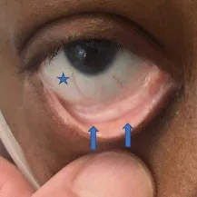
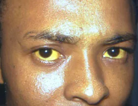
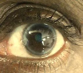
Conjunctival Pallor4 · Scleral Icterus5 · Arcus Senillis6
Report: I note no conjunctival pallor, scleral icterus (jaundice) or arcus senilis (grey arc around cornea).
Why Check: Conjunctival pallor is a sign of anemia; Scleral Icterus (jaundice) can be a sign of Pre-hepatic (e.g. hemolytic anemia), Hepatic (e.g. Acute hepatitis) or Post-Hepatic (obstructive – cholecystitis, choledocholithiasis, etc.) hyperbilirubinemia. Arcus senilis can be a sign of dyslipidemia (lipid deposits)
Hands & Mouth
AFMC level Pre-clerkship 4 · Clerkship 4
Say: I will inspect under the tongue and the hands.
Do: Look for the following abnormalities in the hands and mouth
- Splinter Hemorrhages: tiny blood clots running vertically under nails.
- Janeway Lesions: small nodular lesions on palms
- Osler Nodes: painful red, raised lesions on hands and feet.
- Respiratory findings: clubbing, tar staining, peripheral cyanosis (Ch 5 – Respiratory Exam).
- Capillary Refill Time: <2 seconds for children and adult men; <3 seconds for women; <4 seconds for elderly patients (see Ch 8- Peripheral Vascular Exam).
- Tremor: an involuntary, oscillating movement of a fixed frequency/rhythmic quality.
- Pallor: Assess hands at palmar creases & nailbeds.
- Central Cyanosis: in the mouth, assess under tongue for central cyanosis (see Ch 5 – Respiratory Exam)
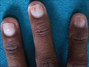
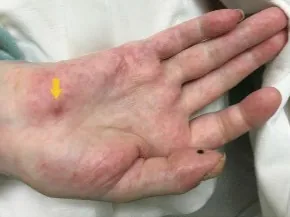
Splinter Hemorrhages7 · Janeway Lesions8
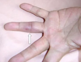
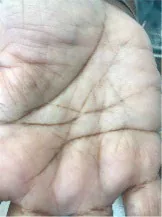
Report: I note no signs of central cyanosis or findings in the hands (in an OSCE station, verbalize all findings).
Why Check:
- Splinter hemorrhages, Janeway lesions and Osler nodes are signs of infective endocarditis due to immune complex deposition and/or septic micro-emboli, and can be used as minor criteria in the Duke Criteria for Infective Endocarditis.
- Tremor can be seen in thyrotoxicosis (see Thyroid Exam – Ch 15 – MF3).
- Pallor in palmar creases & nailbeds are signs of anemia.
- Peripheral cyanosis indicates circulatory insufficiency; central cyanosis indicates poor oxygenation in the lungs (specifically PaO2 <50mmHg given an Hb of 140-160g/L; PaO2 45mmHg if Hb 120 g/L).
Jugular Venous Pressure (JVP) – mix of inspection & palpation
Also seen in Ch 9 – Volume Status Exam – MF2
- JVP - [4, 4]
- JVP Waveform- [2, 3]
Say: Please lay back on the exam table and tilt the head all the way to the left.
How do you differentiate the JVP from the Carotid Pulse?
| JVP | Carotid |
|---|---|
| Occludable and bounces back after occlusion | Non-occludable |
| Non-palpable | Palpable |
| Biphasic | 1-1 Pulse to Heartbeat |
| Decreases: with elevated position and with inspiration | No change with position or inspiration |
| Increases: with Abdomino-Jugular Reflux (See below) | No change with AJR maneuver |
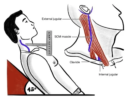
Do:
- Prop up head of exam table to 30-45°. Stand at right side. Look for JVP between 2 heads of sternocleidomastoid (i.e. sternal head medially, clavicular head laterally), or look lateral to sternal head of sternocleidomastoid.
- Once you have located the JVP, measure by placing ruler on sternal angle facing vertically
- Make a right-angle triangle going from the apex of the JVP to the ruler.
- The height on the ruler is the measured JVP.
Note: on OSCE, estimate using your fingers, < 3 finger breadths is normal – not expected to use ruler.
What is Normal: 3-4 cm Above Sternal Angle (ASA)
Report: JVP was located and is < 5 cm above the sternal angle
Why Check: The Jugular Venous Pressure (JVP) is used to check the intravascular volume status of patients (termed “wet” if the patient is volume overloaded and the JVP is elevated, or “dry” if the patient is volume depleted and the JVP is flat). The JVP is an indirect measure of right atrial pressure via visualization of the internal jugular vein. The Central Venous Pressure (cm H2O) can be estimated from the JVP by adding 5cm to the measured value, since right atrium is approximately 5cm below the sternal angle. In the appropriate clinical context (e.g. dyspneic patients in Emergency Department), an elevated JVP increases the likelihood of suspected Congestive Heart Failure (LR+ 5.1; LR- 0.66) If the JVP increases with inspiration, this is called Kussmaul’s Sign and may be a sign of severe pericarditis or right-sided heart failure. Beck’s Triad (for Acute Cardiac Tamponade) includes distended neck veins, low Arterial Blood Pressure, and Muffled Heart Sounds.
Abdominal
AFMC level Pre-clerkship 4 · Clerkship 4
Say: I will briefly inspect the abdomen for abnormalities.
Do: Look for signs of ascites, any pulsations or surgical scars in abdominal area
Report: I note no ascites, pulsations or surgical scars in the abdomen.
Why Check: Looking for signs of portal hypertension – note that patients may have ascites without physical findings (Ultrasound more sensitive). May also look for other signs of venous congestion (e.g. Edema, Jugular Venous Distension – see JVP assessment above), and for other signs of liver disease (e.g. jaundice, coagulopathy, telangiectasias, etc. – see Abdominal Exam – Ch 13 – MF3)
Legs
AFMC level Pre-clerkship 4 · Clerkship 4
Say: I will briefly inspect the legs for abnormalities.
Do: Look for signs of edema, erythema, surgical scars and peripheral vascular disease.
Report: I note no edema, erythema, surgical scars or concerns for peripheral vascular disease.
Why Check: Assessing extent of vasculopathy (see Peripheral Vascular Exam – Ch 8 – MF1 for more details), potential extent of Congestive Heart Failure, etc.
Xanthoma
Say: I will inspect for any visible yellow depositions.
Do: Assess for Xanthomata (yellowish cholesterol-rich fat depositions). Common sites include eyes, joints, hands, and tendons (e.g. Achilles tendon)
Report: No xanthomata visible on inspection
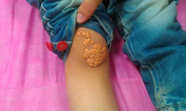
Xanthoma10
Why Check: Xanthomata are complications of hyperlipidemia – significant risk factor of cardiovascular disease
Coagulopathy
AFMC level Pre-clerkship 3 · Clerkship 4
Say: I am going to inspect for any signs of coagulopathy
Do: Assess for Hematomas: Based on size, either Petechiae (1-2 mm diameter red spot), Purpura (3-10 mm), and/or Ecchymosis (>10 mm); as well as Telangiectasias (spider veins)
Report: No Petechiae, purpura or ecchymosis noted on inspection.

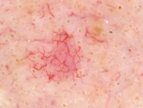
Petechiae11 · Telangiectasias12
Why Check: Signs of both platelet disorders, and vascular disorders.
Palpation
Abdomino-jugular Reflux (AJR) – formerly Hepatojugular Reflux (HJR)
AFMC level Pre-clerkship 2 · Clerkship 3
Say: Please lay back on the exam table and tilt the head to the left. I’m going to press on the upper abdomen and monitor the veins in the neck.
Do: Press firmly over right upper quadrant or centre of abdomen for 10-15 seconds. Observe the JVP. If JVP does rise significantly (≥4cm), observe if JVP returns to baseline while applying pressure. Sustained elevation of JVP beyond 10 seconds is abnormal. This can be confirmed by suddenly releasing abdominal pressure and seeing a return to baseline (≥4cm fall).
What is Normal: In an otherwise healthy individual, the JVP remains constant or temporarily rises for 1-2 heartbeats before returning to normal.
Report: Abdominojugular Reflux lasted less than 10 seconds, which is normal.
Why Check: An alternative test for measuring JVP, method to assess right ventricular failure. This is also useful if you are having trouble seeing or differentiating the JVP from carotid.
Heaves and Thrills
AFMC level Pre-clerkship 1 · Clerkship 2
Say: I will now be palpating the chest at the four valve regions of the heart.
Where to Palpate: See image
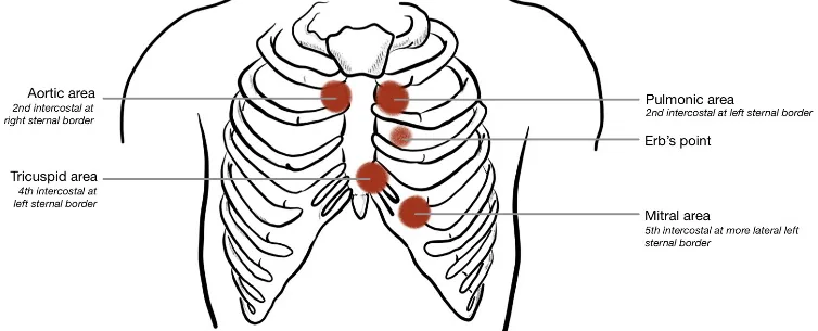
Do: Use whole hand to palpate heaves, then pads of 2-3 fingers to palpate for thrills at each valve region (Heaves = Hands, Thrills = Tips)
Report: There are no heaves or thrills on palpation.
Why Check: Heaves provide evidence of hypertrophy. Thrills provide evidence of turbulent blood flow, complemented by murmurs heard on auscultation.
PMI (Point of Maximal Impulse)
- Loc.– [3, 4]
- Amp. – [1, 1]
- Dur. – [1, 2]
- Size – [2, 4]
Say: I will palpate the chest for where the heart beats strongest. Can you lean forward?
Do: Using 2-3 fingers, locate the 5th intercostal space at left mid-clavicular line (MCL) – the expected site of PMI. Assess Location, Amplitude, Duration and Size (LADS).
Report: PMI located at 5th intercostal at left MCL, is brisk and tapping, lasts 2/3rd systole, and size of quarter.
Why Check: Apex beat being palpated can provide evidence of hypertrophy – PMI not always apical impulse
- Location of Apex: Displaced location can be evidence of decreased ejection fraction, left ventricular dilatation, increased left atrial pressure, or enlarged Cardiothoracic Ratio on Chest X-Ray.
- Amplitude of Apex: If patient has known mitral stenosis, increased apex amplitude suggests comorbid aortic stenosis or mitral regurgitation
- Duration of Apex: Sustained apex with an aortic murmur argues for aortic stenosis; Lack of sustained apex with Aortic Regurgitation argues against moderate-severe severity of Aortic regurgitation
- Size of Apex: Increased size evidence of increased LV End-Diastolic Volume
Edema
AFMC level Pre-clerkship 4 · Clerkship 4
(Also seen in Ch 8 – Peripheral Vascular Exam – MF1)
Say: I will palpate for leg swelling (Sacral edema if patient immobile). Please tell me if you are in discomfort.
Do: Press firmly with pads of your thumbs on the dorsal surface of patient’s leg, just above the ankle at bony prominence. Hold for 10s, then release. Look for pitting edema – skin does not bounce back immediately (i.e. pits). How high does it go on the leg? Is it pitting (transudate) or non-pitting (exudate)? Look at patient for signs of discomfort and stop exam signs of distress.
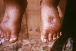
Pedal Edema13
Grading: 0 = none; 1+ = 2mm depression; 2+ = 4mm depression; 3+ = 6mm depression; 4+ = 8mm depression
Report: I note (grade) pitting edema bilaterally, up to the level of (location e.g. ankles).
Why Check: Edema is excess interstitial fluid accumulation causing swelling, particularly in gravity dependent areas (e.g. feet). Normally, this fluid returns through lymphatic drainage. Pitting edema = transudate (non-protein filled) – sign of increased hydrostatic pressure (e.g. congestive heart failure, decreased venous return) or decreased oncotic pressure (e.g. nephrotic syndrome, hypoalbuminemia). Non-pitting edema = exudate (protein) filled, or an issue with lymphatic drainage (e.g. lymphedema).
Central & Upper Limb Pulses
AFMC level Pre-clerkship 1 · Clerkship 2
Say: I will palpate the central pulses in the neck, arms and groin.
Do: Palpate for pulses bilaterally. For peripheral pulses, you can palpate the most distal pulse. If it is non-palpable, move more proximally. Should be symmetrical, regular rate/rhythm and no delay between them. Note that Lower Limb Peripheral Pulses are explored in Ch 8 – Peripheral Vascular Exam.
Where to Palpate – Upper Extremities:
- Radial: Along radius bone just proximal to wrist on palmar side of hand
- Brachial: Medial to biceps tendon and 2-3cm above antecubital fossa
Where to Palpate – Central Pulses:
- Carotid: Medial to sternocleidomastoid muscle, roughly at level of cricoid cartilage
- Abdominal Aorta: Slightly left and superior to umbilicus
- Femoral: Midway between ASIS and Symphysis pubis
Note: Auscultate for carotid bruits before palpation of carotid pulses using bell of stethoscope (see below)
Grading: 0 = Non-palpable; 1 = Barely palpable; 2 = Easily Palpable (Normal); 3 = Bounding; 4 = Aneurysmal.
Report: Central and most distal upper limb peripheral pulses are symmetrical with regular rate and rhythm.
Why Check: Note that palpation of peripheral and central pulses is a core element of the Peripheral Vascular Exam (see Chapter 8 – Peripheral Vascular Exam). Specific findings include Pulses alternans (two strong systolic with mid-systolic dip) suggests LV dysfunction or Cardiac tamponade. An Irregularly irregular rhythm may suggest Atrial fibrillation.
Modification for Patients with Obesity: Use a handheld Doppler Ultrasound to locate pulses if they are difficult to appreciate by palpation. In this population, a doppler may be more sensitive than palpation.
Auscultation
Cardiac Precordial Auscultation
- S3, S4- [1, 3]
- Murmur [1, 3]
- Abnormal S1, S2 [1, 1]
- Position [1, 4]
Say: I will auscultate the chest for heart sounds. Please breathe normally as I listen.
Do: Listen carefully with the diaphragm of the stethoscope at each valve region (Aortic, Pulmonic, Tricuspid, and Mitral). Listen for abnormal heart sounds or murmurs.
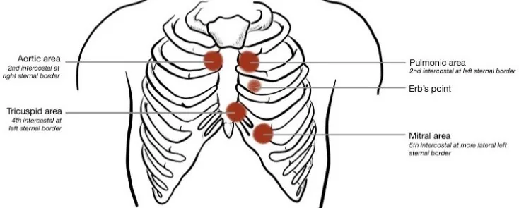
Report: I note a normal S1 and S2. There are no extra heart sounds or murmurs present.
What Is Normal? S1 and S2, regular rate and rhythm, with no extra heart sounds and no murmurs
- S1 = loudest at Tricuspid & Mitral valves (re atrioventricular valves closing at start of systole)
- S2 = loudest at Aortic & Pulmonic valves (re semilunar valves closing at end of systole). Physiologic splitting of S2 is normal on inspiration due to ↑ negative intrathoracic pressure – best heard in a sitting position.
Abnormal Findings – Abnormal S1 and S2 and Associated Conditions:
| Characteristic | S1 | S2 |
|---|---|---|
| Loud | Mitral Valve Prolapse (MVP); Mitral Stenosis (MS); Left Atrial Myxoma; Tachycardia | Systemic hypertension (loud A2) or Pulmonary hypertension (loud P2) |
| Faint | Mitral Regurgitation; Left Bundle Branch Block (LBBB); Long PR interval (1st degree AV block); Acute Aortic Regurgitation (AR) | Aortic Stenosis (LR+ 7.5, LR- 0.5) |
| Variable | AV Dissociation (LR+ 24.4, LR- 0.4); Atrial Fibrillation | N/A |
| Wide Split | Right Bundle Branch Block (RBBB); Right Atrial Myxoma | RBBB; Pulmonic Stenosis; Mitral Regurgitation; Ventricular Septal Defect (VSD); Right Ventricular Failure |
| Paradoxical Split | N/A | LBBB; Severe Aortic Stenosis; Wolff-Parkinson-White (WPW); LV Dysfunction |
| Fixed Split | N/A | Atrial Septal Defect (ASD) |
Abnormal Findings – Extra heart sounds: Best heard in left lateral decubitus position.
- S3: Heard just after S2, with rapid and early diastolic filling of ventricles, “Sloshing in” (e.g. volume overload). In the appropriate clinical context (e.g. dyspneic patients in Emergency Department), hearing an S3 increases the likelihood of Congestive Heart Failure (LR+11, LR- 0.88).
- S4: Heard just before S1, with diastolic rapid late filling of ventricles, “A stiff wall” (e.g. pressure overload, Left Ventricular Hypertrophy).
- S3 + S4: Summation gallop
Abnormal Findings – Murmurs (represent Turbulent blood flow). Listen for Location (i.e. identify valve); Radiation (e.g. to carotids, left sternal border, etc.); Timing (e.g. Systolic vs. Diastolic); Configuration (e.g. Crescendo, Decrescendo, Crescendo-decrescendo, Plateau); Quality (e.g. high vs. low pitch; harsh, musical, blowing, rumbling, etc.); Change with maneuvers (e.g. with inspiration, Valsalva, squat); and Volume/Grade
- Systolic Murmur: Grades 1 (very faint); Grade 2 (soft, readily detectable); Grade 3 (easily detectable without thrill); Grade 4 (easily detectable with thrill); Grade 5 (very loud murmur audible with stethoscope placed lightly on chest); Grade 6 (extremely loud, audible with stethoscope off chest)
- Diastolic Grade: Grade 1 (barely audible); Grade 2 (audible, soft); Grade 3 (easily audible); Grade 4 (loud)
Mitral Stenosis (MS)
- Pathophysiology: Mitral valve should be open in diastole – mitral stenosis causes incomplete opening of mitral valve in diastole
- Murmur: Diastolic murmur with Opening Snap best heard at cardiac apex in left lateral decubitus position. Worsened by ↑ venous return (squat/leg raise)
Aortic Regurgitation / Insufficiency (AR/AI)
- Pathophysiology: Aortic valve should be closed in diastole – aortic regurgitation causes incomplete closing of aortic valve in diastole
- Murmur: Diastolic murmur heard best at right base in sitting position. Worsened by ↑ venous return (squat/leg raise)
Aortic Stenosis (AS)
- Pathophysiology: Aortic valve should be open in systole – aortic stenosis causes incomplete opening of aortic valve in systole
- Murmur: Systolic murmur with crescendo-decrescendo pattern heard best at right base. Radiates to R clavicle and/or R carotid. Worsened by ↑ venous return (squat/leg raise)
Hypertrophic Cardiomyopathy (HCM)
- Pathophysiology: LV Outflow Tract Obstruction due to sarcomere gene mutations.
- Murmur: Systolic murmur with crescendo-decrescendo pattern heard best at cardiac apex and left lower sternal border. May radiate to axilla. Improved by ↑ venous return (squat/leg raise)
Mitral Regurgitation (MR)
- Pathophysiology: Mitral valve should be closed in systole – mitral regurgitation causes incomplete closing of mitral valve in systole.
- Murmur: Systolic murmur with holosystolic pattern heard best at cardiac apex. Radiates to axilla. Worsened by ↑ venous return (squat/leg raise)
Mitral Valve Prolapse (MVP)
- Pathophysiology: Mitral valve should be closed in systole, where thickened mitral valve leaflets in MVP cause imperfect closure of mitral valve in systole.
- Murmur: Systolic murmur similar to Mitral regurgitation, but improved by ↑ venous return (squat/leg raise)
Maneuvers to Accentuate/Differentiate Murmurs:
| Maneuver | RV Preload | LV Preload | LV Afterload | Effect on Murmurs |
|---|---|---|---|---|
| Inspiration | ↑ | ↓ | ∅ | ↑ R-sided murmurs, ↓ L-sided murmurs (except HCM) |
| Valsalva (Strain Phase)/ Abrupt Stand | ↓ | ↓ | ↓ | ↑ MVP/HCM, ↓ MS/MR/AS/AR/VSD |
| Squatting/Leg raise | ↑ | ↑ | ∅/↑ | ↓ MVP/HCM, ↑ MS/MR/AS/AR/VSD |
| Hand Grip (for 20-30s) | ∅ | ∅ | ↑ | ↓ MVP/HCM; ↑ MR/AR/VSD; ↓ MS/AS |
Note on Pediatric Cardiac Auscultation: Not all pediatric murmurs are pathologic; pathology more likely if murmur is systolic grade 3+; holosystolic; associated systolic click; diastolic; worse on standing; or harsh.
Modification for Patients with Obesity: Ask the patient to lean forward if sitting, or to place their above their head if supine. Leaning forward brings the heart closer to the chest wall & placing arms above the head when supine redistributes the soft tissue of the chest wall, making heart sounds easier to appreciate.
Carotid Bruits
AFMC level Pre-clerkship 2 · Clerkship 3
Say: I will auscultate the carotid arteries in the neck (point on yourself to where you will auscultate) for signs of turbulent blood flow.
Do: Listen with Bell of Stethoscope. Listen for duration, intensity, and pitch. If applicable, also listen for any radiation of a systolic murmur to the right carotid (e.g. Aortic Stenosis)
Report: I note no carotid bruits on auscultation bilaterally.
Why Check: Can assess for carotid artery stenosis prior to palpating, where the presence of a carotid bruit has a 53% sensitivity and 83% specificity of a clinically relevant stenosis14. In addition, radiation of a systolic murmur to the right carotid ↑ likelihood of suspected aortic stenosis (LR+ 8.1, LR- 0.29)
Carotid Upstroke – Conditional Test
AFMC level Pre-clerkship 2 · Clerkship 3
Say: I will continue auscultating the heart while I also palpate over a carotid artery on one side of the neck (point on yourself to where you will palpate).
Setup: Auscultate over the valve that you suspect may be stenotic or regurgitant. Place the pads of your 2-3 fingers over the carotid artery gently (after auscultating for carotid bruits).
Do: As you listen for murmur, there should be no delay between murmur onset (systole) and carotid pulse.
- If carotid pulse is delayed and relatively diminished, this is Pulsus parvis et tardus (sign of aortic stenosis).
- If the carotid pulse is brisk, this may be a sign of aortic regurgitation.
- If the carotid pulse is biphasic (2 pulses per cardiac cycle), this is called pulsus bisferiens and may be a sign of coexisting Aortic Stenosis and Regurgitation.
Report: I note a normal carotid upstroke with no delay between the murmur and carotid pulse.
Why Check: Carotid upstroke is a conditional test that can be used if you suspect an aortic stenosis or aortic regurgitation murmur. Aortic stenosis is expected to cause carotid upstroke to be delayed and diminished due to partially obstructed forward flow, where this finding (pulsus parvis et tardus) increases likelihood of suspected aortic stenosis (LR + 9.2, LR- 0.56). Aortic regurgitation is expected to cause brisk carotid upstroke due to ↑ preload from regurgitation. Biphasic carotid pulse (pulsus bisferiens) due to comorbid Aortic stenosis and Aortic regurgitation is a combination of these morphologies.
Helpful Stethopedia Videos to Review
- Video 1 – Cardiac Exam OSCE Style (Link: https://videopress.com/v/k7npdLgU)
- Video 2 – Cardiac Exam Special Tests (Link: https://videopress.com/v/FvzO3Mim https://videopress.com/v/PBR0adsa)
Part 4: Relevant Likelihood Ratios – from Rational Clinical Examination JAMA series
Clinical Question 1: Is this patient having a Myocardial Infarction?1516
Table 1: Likelihood Ratios of Chest Pain Radiation Patterns for Myocardial Infarction15
| Procedure | LR+ (95% CI) | LR- (95% CI) |
|---|---|---|
| Both arms with pain | 9.7 (4.6-20) (about +30%) | 0.64 (0.54-0.74) (about −15%) |
| Right arm pain | 7.3 (3.9-14) (about +30%) | 0.62 (0.52-0.73) (about −15%) |
| Left arm pain | 2.2 (1.6-301) (about +15%) | 0.60 (0.48-0.75) (about −15%) |
| Right shoulder pain | 2.2 (1.4-3.4) (about +15%) | 0.90 (0.82-0.97) (about −15%) |
Table 2: Acute MI in patients with Undifferentiated Chest Pain Admitted for Suspected Acute Coronary Syndrome16
| Procedure | LR+ (95% CI) | LR- (95% CI) |
|---|---|---|
| Radiation to the shoulder or both arms | 4.1 (2.5-6.5) (about +15%) | 0.68 (0.52-0.89) (about −15%) |
| Radiation to right arm | 3.8 (2.2-6.6) (about +15%) | 0.86 (0.77-0.96) (about −15%) |
| Vomiting | 3.5 (2.0-6.2) (about +15%) | 0.87 (0.79-0.97) (about −15%) |
| Ex-smoker | 2.5 (1.6-4.0) (about +15%) | 0.85 (0.76-0.96) (about −15%) |
| Male sex | 1.5 (1.3-1.6) (about +15%) | 0.24 (0.12-0.48) (about −30%) |
| Current smoker | 1.4 (1.0-1.8) (no change: 95% CI includes 1) | 0.83 (0.68-1.0) (no change: 95% CI includes 1) |
| Radiation to left arm | 1.3 (0.93-1.8) (no change: 95% CI includes 1) | 0.90 (0.76-1.1) (no change: 95% CI includes 1) |
Table 3: Pre-Test Likelihood of Coronary Artery Disease using Chest Pain Criteria16:
| Age (Years) | Non-anginal chest pain 1 of 3 chest pain criteria Male | Non-anginal chest pain 1 of 3 chest pain criteria Female | Atypical angina 2 of 3 chest pain criteria Male | Atypical angina 2 of 3 chest pain criteria Female | Typical angina 3 of 3 chest pain criteria Male | Typical angina 3 of 3 chest pain criteria Female |
|---|---|---|---|---|---|---|
| 30-39 | 4% | 2% | 34% | 12% | 76% | 26% |
| 40-49 | 13% | 3% | 51% | 22% | 87% | 55% |
| 50-59 | 20% | 7% | 65% | 33% | 93% | 73% |
| 60-69 | 27% | 14% | 72% | 51% | 94% | 86% |
Note – 3 Chest Pain Criteria:
- Substernal chest discomfort with characteristic quality and duration
- Provoked by exertion or emotional stress
- Relieved promptly by rest or nitroglycerin
Clinical Question 2: Does this patient have an Abnormal Systolic Murmur?1718
Table 1: Likelihood Ratios of Individual Findings for Identifying Systolic Murmurs that are Significant (Moderate-to-Severe Aortic Stenosis or Mitral Regurgitation, Congenital Shunt, or Intraventricular Pressure Gradient)17
| Procedure | LR+ (95% CI) | LR- (95% CI) |
|---|---|---|
| Systolic Thrill | 12 (0.76-205) (no change: 95% CI includes 1) | 0.73 (0.58-0.93) (about −15%) |
| Holosystolic Murmur | 8.7 (2.3-33) (about +30%) | 0.19 (0.08-0.43) (about −30%) |
| Loud Murmur | 6.5 (2.3-19) (about +30%) | 0.08 (0.02-0.31) (about −45%) |
| Plateau-Shaped Murmur | 4.1 (1.4-12) (about +15%) | 0.48 (0.30-0.77) (about −30%) |
| Loudest at the Apex | 2.5 (0.58-11) (no change: 95% CI includes 1) | 0.84 (0.65-1.1) (no change: 95% CI includes 1) |
| Radiation to the Carotid | 0.91 (0.28-3.0) (no change: 95% CI includes 1) | 1.0 (0.78-1.3) (no change: 95% CI includes 1) |
Table 2: Accuracy of Physical Examination for Detecting Moderate or Severe Aortic Stenosis17
| Procedure | LR (95% CI) |
|---|---|
| Systolic murmur over right clavicle and 3-4 associated findings | 40 (6.6-239) |
| Systolic murmur over right clavicle and 0-2 associated findings | 1.8 (0.93-2.9) |
| No systolic murmur over right clavicle | 0.1 (0.02-0.44) |
- Associated Findings:
- Reduced or absent second heart sound (S2)
- Reduced carotid volume
- Slow rate of increase of carotid pulse (slow carotid upstroke)
- Maximal murmur intensity in second right intercostal space
- Note: Moderate or severe aortic stenosis defined as valve area <1.2cm² or peak transvalvular gradient ≥25mmHg
Table 3: Accuracy of Physical Examination for Detecting Severe Mitral Regurgitation17
| Procedure | LR+ (95% CI) |
|---|---|
| Murmur Grades 4-5 | 14 (3.3-56) (about +45%) |
| Murmur Grade 3 | 3.5 (2.1-5.7) (about +15%) |
| Murmur Grades 0-2 | 0.19 (0.11-0.33) (about −30%) |
- Note: Severe mitral regurgitation defined as regurgitant fraction of 40% or more detected by echocardiography
Table 4: Accuracy of Physical Examination for Detecting Tricuspid Regurgitation18
| Procedure | LR+ (95% CI) | LR- (95% CI) |
|---|---|---|
| Characteristic Murmur (mild or worse Tricuspid regurgitation) | 14.6 (4.5-47.1) (about +45%) | 0.8 (0.7-0.9) (about −15%) |
| Characteristic Murmur (moderate-to-severe Tricuspid regurgitation) | 10.1 (5.8-17.8) (about +45%) | 0.4 (0.2-0.7) (about −30%) |
| Pulsatile Liver (moderate-to-severe) | 3.9 (1.4-11.4) (about +15%) | 0.8 (0.6-1.0) (no change: 95% CI includes 1) |
Clinical Question 3: Does this patient with a Pericardial Effusion have Cardiac Tamponade?19
| Procedure | Sensitivity | Specificity | LR+ (95% CI) | LR- (95% CI) |
|---|---|---|---|---|
| Pulsus Paradoxus > 12 mmHg | 0.98 | 0.83 | 5.9 (2.4-14) (about +30%) | 0.03 (0-0.21) (about −45%) |
| Pulsus Paradoxus > 10 mmHg | 0.98 | 0.70 | 3.3 (1.8-6.3) (about +15%) | 0.03 (0.01-0.24) (about −45%) |
Clinical Question 4: Does this dyspneic patient in the Emergency Department have Congestive Heart Failure?20
| Procedure | LR+ (95% CI) | LR- (95% CI) |
|---|---|---|
| History: Patient History of Heart Failure | 5.8 (4.1-8.0) (about +30%) | 0.45 (0.38-0.53) (about −30%) |
| History: Patient History of Myocardial Infarction | 3.1 (2.0-4.9) (about +15%) | 0.69 (0.58-0.82) (about −15%) |
| Physical Exam: Third Heart Sound (S3) | 11 (4.9-25) (about +45%) | 0.88 (0.83-0.94) (about −15%) |
| Physical Exam: Jugular Venous Distension | 5.1 (3.2-7.9) (about +30%) | 0.66 (0.57-0.77) (about −15%) |
| Physical Exam: Rales | 2.8 (1.9-4.1) (about +15%) | 0.51 (0.37-0.70) (about −15%) |
| Physical Exam: Abdominojugular Reflux | 6.4 (0.81-51) (no change: 95% CI includes 1) | 0.79 (0.62-1.0) (no change: 95% CI includes 1) |
| Chest X-Ray: Pulmonary Venous Congestion | 12 (6.8-21) (about +45%) | 0.48 (0.28-0.83) (about −30%) |
| Chest X-Ray: Interstitial Edema | 12 (5.2-27) (about +45%) | 0.68 (0.54-0.85) (about −15%) |
| Chest X-Ray: Alveolar Edema | 6.0 (2.2-16) (about +30%) | 0.95 (0.93-0.97) (about −15%) |
| Chest X-Ray: Cardiomegaly | 3.3 (2.4-4.7) (about +15%) | 0.33 (0.23-0.48) (about −30%) |
| Chest X-Ray: Pleural Effusion(s) | 3.2 (2.4-4.3) (about +15%) | 0.81 (0.77-0.85) (about −15%) |
| ECG: Atrial Fibrillation | 3.8 (1.7-8.8) (about +15%) | 0.79 (0.65-0.96) (about −15%) |
| ECG: New T-Wave Changes | 3.0 (1.7-5.3) (about +15%) | 0.83 (0.74-0.92) (about −15%) |
| ECG: Any Abnormal Finding | 2.2 (1.6-3.1) (about +15%) | 0.64 (0.47-0.88) (about −15%) |
| B-Type Natriuretic Peptide (BNP) pg/mL ≥ 250 | 4.6 (2.6-8.0) (about +15%) | N/A |
| B-Type Natriuretic Peptide (BNP) pg/mL ≥ 100 | 2.7 (2.0-3.9) (about +15%) | N/A |
| B-Type Natriuretic Peptide (BNP) pg/mL ≥ 50 | 1.7 (1.2-2.6) (about +15%) | N/A |
| B-Type Natriuretic Peptide (BNP) pg/mL < 50 | 0.06 (0.03-0.12) (about −45%) | N/A |
| Overall Clinical Impression: Initial clinical judgment that the patient is in heart failure | 4.4 (1.8-10) (about +15%) | 0.45 (0.28-0.73) (about −30%) |
Clinical Question 5: Does this patient have Atrioventricular Dissociation?18
| Procedure | LR+ (95% CI) | LR- (95% CI) |
|---|---|---|
| Changing intensity of S1 | 24.4 (1.5-384.5) (about +45%) | 0.4 (0.3-0.7) (about −30%) |
| Intermittent cannon A waves | 3.8 (1.8-8.2) (about +15%) | 0.1 (0-0.4) (about −45%) |
| Varying arterial pulse | 2.1 (1.0-4.4) (no change: 95% CI includes 1) | 0.5 (0.3-1.0) (no change: 95% CI includes 1) |
Clinical Question 6: Does this patient have an Aortic Dissection?18
| Procedure | LR+ (95% CI) | LR- (95% CI) |
|---|---|---|
| Focal Neurologic Signs | 33.4 (2.0-549) (about +45%) | 0.9 (0.8-1.0) (no change: 95% CI includes 1) |
| Pulse Deficit | 6.0 (1.1-32.5) (about +30%) | 0.7 (0.5-1.0) (no change: 95% CI includes 1) |
| Aortic Regurgitation Murmur | 1.4 (0.9-2.2) (no change: 95% CI includes 1) | 0.9 (0.8-1.0) (no change: 95% CI includes 1) |
| 3 of 3 Above Signs | 65.8 (4.1-1062) (about +45%) | N/A |
| 2 of 3 Above Signs | 5.3 (3.0-9.4) (about +30%) | N/A |
| 1 of 3 Above Signs | 0.5 (0.4-0.8) (about −15%) | N/A |
| 0 of 3 Above Signs | 0.1 (0-0.2) (about −45%) | N/A |
Clinical Question 7: Does this patient have Hypertrophic Cardiomyopathy?18
| Procedure | LR+ (95% CI) | LR- (95% CI) |
|---|---|---|
| Systolic murmur louder with Valsalva maneuver | 14.0 (3.4-57.4) (about +45%) | 0.3 (0.1-0.8) (about −30%) |
| Systolic murmur Softer with Passive leg elevation | 9.0 (3.5-23.3) (about +30%) | 0.1 (0-0.7) (about −45%) |
| Systolic murmur Softer with Standing-to-squatting | 7.6 (2.5-22.7) (about +30%) | 0.1 (0-0.4) (about −45%) |
| Systolic murmur louder with Squatting-to-standing | 6.0 (2.9-12.3) (about +30%) | 0.1 (0-0.8) (about −45%) |
| Systolic murmur Softer with Isometric hand grip | 3.6 (2.0-6.4) (about +15%) | 0.1 (0-0.9) (about −45%) |
Clinical Question 8: Does this patient have Anemia?18
Note: Anemia defined as Hct <35%, Hgb <110g/dL, or Hgb <11g/dL for women and <13 g/dL for men
| Procedure | LR+ (95% CI) | LR- (95% CI) |
|---|---|---|
| Conjunctival Pallor Rim Present | 16.7 (2.2-124.7) (about +45%) | N/A |
| Palmar Crease Pallor | 7.9 (1.8-35.3) (about +30%) | 0.9 (0.9-1.0) (no change: 95% CI includes 1) |
| Palmar Pallor | 5.6 (1.1-29.1) (about +30%) | 0.4 (0.4-0.5) (about −30%) |
| Conjunctival Pallor | 4.7 (1.9-11.5) (about +15%) | 0.6 (0.4-0.9) (about −15%) |
| Pallor at any site | 4.1 (2.0-8.7) (about +15%) | 0.4 (0.3-0.7) (about −30%) |
| Nailbed Pallor | 3.9 (0.8-18.6) (no change: 95% CI includes 1) | 0.5 (0.4-0.7) (about −15%) |
| Facial Pallor | 3.8 (2.5-5.8) (about +15%) | 0.6 (0.5-0.7) (about −15%) |
| Conjunctival Pallor Rim Borderline | 2.3 (1.5-3.5) (about +15%) | N/A |
| Conjunctival Pallor Rim Absent | 0.6 (0.5-0.8) (about −15%) | N/A |
Clinical Question 9: Does this patient with Palpitations have a Significant Cardiac Arrhythmia?21
| History | LR+ (95% CI) | LR- (95% CI) |
|---|---|---|
| Regular rapid-pounding sensation in neck | 177 (25-1251) (about +45%) | 0.07 (0.03-0.19) (about −45%) |
| Visible neck pulsations | 2.68 (1.25-5.78) (about +15%) | 0.87 (0.76-1.00) (no change: 95% CI includes 1) |
| Affected by Sleeping | 2.44 (1.43-4.14) (about +15%) | 0.63 (0.42-0.93) (about −15%) |
| Age >60 years old | 1.89 (1.03-3.47) (about +15%) | 0.77 (0.56-1.06) (no change: 95% CI includes 1) |
| Vasovagal Symptoms (pale and/or sweaty) | 1.72 (1.11-2.65) (about +15%) | 0.63 (0.39-1.03) (no change: 95% CI includes 1) |
| Palpitations Character: Regular | 1.38 (1.02-1.86) (about +15%) | 0.55 (0.27-1.14) (no change: 95% CI includes 1) |
| Affected by Caffeine | 2.06 (0.78-5.39) (no change: 95% CI includes 1) | 0.89 (0.72-1.09) (no change: 95% CI includes 1) |
| Affected by Alcohol | 1.94 (0.74-5.12) (no change: 95% CI includes 1) | 0.90 (0.74-1.09) (no change: 95% CI includes 1) |
| Occurring at work | 1.54 (0.78-3.04) (no change: 95% CI includes 1) | 0.86 (0.65-1.14) (no change: 95% CI includes 1) |
| Dizzy spells | 1.34 (0.96-1.88) (no change: 95% CI includes 1) | 0.67 (0.39-1.18) (no change: 95% CI includes 1) |
| Male Sex | 1.20 (0.69-2.11) (no change: 95% CI includes 1) | 0.90 (0.65-1.26) (no change: 95% CI includes 1) |
| Palpitations Duration: >60s | 1.17 (0.93-1.48) (no change: 95% CI includes 1) | 0.63 (0.27-1.44) (no change: 95% CI includes 1) |
| Heart Rate >100 beats/min | 1.08 (0.78-1.50) (no change: 95% CI includes 1) | 0.86 (0.44-1.68) (no change: 95% CI includes 1) |
| Family History of Palpitations | 1.07 (0.49-2.37) (no change: 95% CI includes 1) | 0.98 (0.78-1.24) (no change: 95% CI includes 1) |
| Presyncope | 1.04 (0.71-1.51) (no change: 95% CI includes 1) | 0.95 (0.57-1.61) (no change: 95% CI includes 1) |
| Occurring while lying in bed | 1.02 (0.73-1.44) (no change: 95% CI includes 1) | 0.97 (0.57-1.65) (no change: 95% CI includes 1) |
| Occurring while resting | 1.02 (0.69-1.50) (no change: 95% CI includes 1) | 0.97 (0.56-1.69) (no change: 95% CI includes 1) |
| Alcohol Use (>10 drinks/week) | 1.02 (0.38-2.79) (no change: 95% CI includes 1) | 1.00 (0.83-1.19) (no change: 95% CI includes 1) |
| Continuous Symptoms | 0.93 (0.71-1.23) (no change: 95% CI includes 1) | 1.20 (0.62-2.31) (no change: 95% CI includes 1) |
| Anxiety Disorder | 0.92 (0.49-1.72) (no change: 95% CI includes 1) | 1.04 (0.78-1.39) (no change: 95% CI includes 1) |
| Chest pain | 0.92 (0.42-1.98) (no change: 95% CI includes 1) | 1.02 (0.81-1.30) (no change: 95% CI includes 1) |
| Neck fullness | 0.85 (0.44-1.64) (no change: 95% CI includes 1) | 1.04 (0.90-1.20) (no change: 95% CI includes 1) |
| Palpitations Duration: >5 min | 0.79 (0.46-1.36) (no change: 95% CI includes 1) | 1.23 (0.81-1.86) (no change: 95% CI includes 1) |
| Occurring during holiday | 0.79 (0.25-2.49) (no change: 95% CI includes 1) | 1.04 (0.88-1.24) (no change: 95% CI includes 1) |
| Affected by Exercise | 0.78 (0.33-1.82) (no change: 95% CI includes 1) | 1.07 (0.87-1.33) (no change: 95% CI includes 1) |
| Smoking (>11 cigarettes/day) | 0.77 (0.26-2.25) (no change: 95% CI includes 1) | 1.03 (0.91-1.17) (no change: 95% CI includes 1) |
| Occurring during weekend | 0.72 (0.27-1.88) (no change: 95% CI includes 1) | 1.08 (0.89-1.32) (no change: 95% CI includes 1) |
| Any Psychiatric Disorder | 0.67 (0.29-1.58) (no change: 95% CI includes 1) | 1.12 (0.91-1.37) (no change: 95% CI includes 1) |
| Affected by Breathing | 0.52 (0.20-1.33) (no change: 95% CI includes 1) | 1.20 (0.98-1.48) (no change: 95% CI includes 1) |
| PMHx Cardiac Disease | 0.42 (0.06-3.06) (no change: 95% CI includes 1) | 1.07 (0.96-1.20) (no change: 95% CI includes 1) |
| Dyspnea | 0.27 (0.04-1.96) (no change: 95% CI includes 1) | 1.12 (1.01-1.25) (about +15%) |
Clinical Question 10: Does this patient in Hypovolemic Shock have a Systolic Blood Pressure >60 mmHg?18
| Procedure | LR+ (95% CI) | LR- (95% CI) |
|---|---|---|
| Radial Pulse Present | 4.7 (0.7-31.3) (no change: 95% CI includes 1) | 0.5 (0.3-0.9) (about −15%) |
| Femoral Pulse Present | 2.9 (1.1-7.2) (about +15%) | 0.1 (0-0.5) (about −45%) |
| Carotid Pulse Present | 1.2 (0.9-1.8) (no change: 95% CI includes 1) | 0.2 (0-2.1) (no change: 95% CI includes 1) |
Clinical Question 11: What is the potential clinical significance of inspection of Neck Veins?18
| Procedure | LR+ (95% CI) | LR- (95% CI) |
|---|---|---|
| Predicting postoperative infarction or death | 11.3 (5.0-25.8) (about +45%) | 0.8 (0.7-1.0) (no change: 95% CI includes 1) |
| Detecting measured CVP > 12cm H2O | 10.4 (5.5-19.9) (about +45%) | 0.1 (0-0.6) (about −45%) |
| Detecting measured CVP >8cm H2O | 9.0 (3.7-22.1) (about +30%) | 0.2 (0-1.5) (no change: 95% CI includes 1) |
| Abdominojugular Reflux, detecting elevated left heart pressures | 8.0 (2.1-31.2) (about +30%) | 0.3 (0.2-0.6) (about −30%) |
| Detecting low LV Ejection Fraction | 7.9 (2.8-22.4) (about +30%) | 0.9 (0.8-1.0) (no change: 95% CI includes 1) |
| Detecting elevated left heart diastolic pressures | 3.9 (1.6-9.4) (about +15%) | 0.7 (0.5-1.0) (no change: 95% CI includes 1) |
Clinical Question 12: What is the potential clinical significance of palpation of the Apical Impulse?18
| Feature | Procedure | LR+ (95% CI) | LR- (95% CI) |
|---|---|---|---|
| Location | Supine apical impulse lateral to midclavicular line (Low ejection fraction) | 10.1 (3.8-26.6) (about +45%) | 0.6 (0.5-0.9) (about −15%) |
| Location | Supine apical impulse lateral to midclavicular line (Increased ventricular volume) | 8.0 (1.9-33.0) (about +30%) | 0.7 (0.6-0.9) (about −15%) |
| Location | Supine apical impulse lateral to midclavicular line (Wedge pressure >12 mmHg) | 5.8 (1.3-26.0) (about +30%) | 0.6 (0.4-1.0) (no change: 95% CI includes 1) |
| Location | Supine apical impulse >10cm from midsternal line (Cardiothoracic ratio >0.5) | 4.3 (0.3-70.8) (no change: 95% CI includes 1) | 0.5 (0.3-0.8) (about −15%) |
| Location | Supine apical impulse lateral to midclavicular line (Cardiothoracic ratio >0.5) | 3.4 (1.6-7.3) (about +15%) | 0.6 (0.5-0.8) (about −15%) |
| Amplitude | Hyperkinetic apical movement (mitral regurgitation or aortic valve disease, patients with mitral stenosis) | 11.2 (6.4-19.5) (about +45%) | 0.3 (0.2-0.4) (about −30%) |
| Duration | Sustained apical movement (severe aortic stenosis in patients with aortic flow murmurs) | 4.1 (1.7-10.1) (about +15%) | 0.3 (0.1-0.5) (about −30%) |
| Duration | Sustained left parasternal movement (RV pressure >50 mmHg) | 3.6 (1.4-8.9) (about +15%) | 0.4 (0.2-0.7) (about −30%) |
| Duration | Sustained apical movement (moderate-to-severe aortic regurgitation in patients with basal early diastolic murmurs) | 2.4 (1.4-4.0) (about +15%) | 0.1 (0-0.9) (about −45%) |
| Size | Apical beat diameter >4cm in lateral decubitus position (Increased LV End-diastolic volume) | 4.7 (2.1-10.2) (about +15%) | 0.4 (0.2-1.0) (no change: 95% CI includes 1) |
Clinical Question 13: What is the potential clinical significance of Abnormal Heart Sounds on Auscultation?18
| Feature | Procedure | LR+ (95% CI) | LR- (95% CI) |
|---|---|---|---|
| S1 | Changing intensity (Atrioventricular dissociation) | 24.4 (1.5-384.5) (about +45%) | 0.4 (0.3-0.7) (about −30%) |
| S2 | Palpable P2 (Pulmonary Hypertension) | 3.6 (1.5-8.8) (about +15%) | 0.05 (0-0.8) (about −45%) |
| S2 | Paradoxical splitting (Significant Aortic Stenosis) | 2.4 (0.8-7.0) (no change: 95% CI includes 1) | 0.6 (0.2-1.7) (no change: 95% CI includes 1) |
| S2 | Fixed wide splitting (Atrial Septal Defect – ASD) | 2.6 (1.6-4.3) (about +15%) | 0.1 (0-0.8) (about −45%) |
| S2 | Loud P2 (Pulmonary Hypertension) | 1.2 (0.9-1.5) (no change: 95% CI includes 1) | 0.8 (0.3-1.9) (no change: 95% CI includes 1) |
| S3 | Predicting postoperative pulmonary edema | 14.6 (5.7-37.3) (about +45%) | 0.8 (0.7-1.0) (no change: 95% CI includes 1) |
| S3 | Detecting elevated BNP levels | 10.1 (4.2-23.9) (about +45%) | 0.5 (0.3-0.8) (about −15%) |
| S3 | Detecting ejection fraction <50% in aortic regurgitation | 8.3 (3.6-19.2) (about +30%) | 0.4 (0.2-0.9) (about −30%) |
| S3 | Predicting postoperative myocardial infarction or death | 8.0 (2.7-23.4) (about +30%) | 0.9 (0.8-1.0) (no change: 95% CI includes 1) |
| S3 | Detecting regurgitant fraction >40% in aortic regurgitation | 5.9 (1.4-25.3) (about +30%) | 0.8 (0.7-0.9) (about −15%) |
| S3 | Detecting elevated left heart filling pressures | 5.7 (3.1-10.3) (about +30%) | 0.8 (0.7-1.0) (no change: 95% CI includes 1) |
| S3 | Detecting ejection fraction <50% in Aortic stenosis | 5.7 (2.7-12.0) (about +30%) | 0.8 (0.7-0.9) (about −15%) |
| S3 | Detecting ejection fraction <30% | 4.1 (2.3-7.3) (about +15%) | 0.3 (0.2-0.5) (about −30%) |
| S3 | Detecting ejection fraction <50% | 3.4 (2.6-4.4) (about +15%) | 0.7 (0.5-0.9) (about −15%) |
| S3 | Detecting myocardial infarction in patients with acute chest pain | 3.2 (1.6-6.5) (about +15%) | 0.9 (0.8-1.0) (no change: 95% CI includes 1) |
| S3 | Detecting pulmonary capillary wedge pressures >12mmHg in Aortic stenosis | 2.3 (1.3-4.0) (about +15%) | 0.9 (0.8-1.0) (no change: 95% CI includes 1) |
| S3 | Detecting depressed ejection fraction in Mitral regurgitation | 1.9 (1.2-2.9) (about +15%) | 0.6 (0.4-0.9) (about −15%) |
| S3 | Detecting elevated left heart filling pressures in Mitral regurgitation | 1.7 (1.0-3.0) (no change: 95% CI includes 1) | 0.7 (0.5-0.9) (about −15%) |
| S4 | Predicting 5-year mortality after infarction | 3.2 (1.3-7.8) (about +15%) | 0.8 (0.6-1.1) (no change: 95% CI includes 1) |
| S4 | Detecting elevated filling pressures | 1.4 (0.8-2.6) (no change: 95% CI includes 1) | 0.6 (0.3-1.4) (no change: 95% CI includes 1) |
| S4 | Detecting severe aortic stenosis | 0.9 (0.5-1.9) (no change: 95% CI includes 1) | 1.1 (0.6-1.9) (no change: 95% CI includes 1) |
Clinical Question 14: Did This Patient Have Cardiac Syncope?22
| Procedure | LR+ (95% CI) | LR- (95% CI) |
|---|---|---|
| Patient Demographics – Atrial fibrillation or flutter | 7.3 (2.4-22) (about +30%) | 0.89 (0.82-0.97) (about −15%) |
| Patient Demographics – Severe structural heart disease | 3.3-4.8 (about +15%) | 0.58-0.70 (about −15%) |
| Patient Demographics – History of heart failure | 2.7 – 3.4 (about +15%) | 0.39 – 0.78 (about −30%) |
| Patient Demographics – Age at first syncopal spell > 35 years old | 3.3 (2.6-4.1) (about +15%) | 0.13 (0.06-0.25) (about −30%) |
| Precipitating or Predisposing Factors – During effort | 1.4 – 15 (about +15%) | 0.88 – 0.96 (about −15%) |
| Precipitating or Predisposing Factors – While supine | 1.1 – 4.9 (about +15%) | 0.89-1.0 (about −15%) |
| Precipitating or Predisposing Factors – Prolonged sitting/standing | 0.54 (0.41-0.72) (about −15%) | 2.0 (1.6-2.6) (about +15%) |
| Precipitating or Predisposing Factors – On the way to the toilet | 0.28 (0.10-0.76) (about −30%) | 1.1 (1.1-1.2) (about +15%) |
| Precipitating or Predisposing Factors – Stress | 0.25 (0.12-0.51) (about −30%) | 1.4 (1.2-1.5) (about +15%) |
| Precipitating or Predisposing Factors – Warm place | 0.17 (0.08-0.33) (about −30%) | 2.0 (1.7-2.4) (about +15%) |
| Precipitating or Predisposing Factors – Pain or medical procedure | 0.12 (0.05-0.28) (about −30%) | 1.8 (1.5-2.1) (about +15%) |
| Precipitating or Predisposing Factors – After using the toilet | 0.05 (0.003 – 0.85) (about −45%) | 1.1 (1.1-1.2) (about +15%) |
| Symptoms Prior to the Episode – Dyspnea | 3.5 (1.5-9.1) (about +15%) | 0.87 (0.74-0.94) (about −15%) |
| Symptoms Prior to the Episode – Chest pain/Angina | 3.4-3.8 (about +15%) | 0.71-0.79 (about −15%) |
| Symptoms Prior to the Episode – Palpitations | 1.9 (0.86-4.5) (no change: 95% CI includes 1) | 0.94 (0.89-1.0) (no change: 95% CI includes 1) |
| Symptoms Prior to the Episode – Absence of prodromes | 1.6 (1.0-2.6) (no change: 95% CI includes 1) | 0.79 (0.69-0.96) (about −15%) |
| Symptoms Prior to the Episode – Pallor | 0.69 (0.58-0.82) (about −15%) | 1.2 (1.0-1.4) (no change: 95% CI includes 1) |
| Symptoms Prior to the Episode – Blurred vision | 0.55 (0.27-1.1) (no change: 95% CI includes 1) | 1.2 (0.96-1.5) (no change: 95% CI includes 1) |
| Symptoms Prior to the Episode – Diaphoresis | 0.49 (0.33-0.71) (about −30%) | 1.2 (1.1-1.3) (about +15%) |
| Symptoms Prior to the Episode – Nausea | 0.44 (0.31-0.62) (about −30%) | 1.1 (1.1-1.3) (about +15%) |
| Symptoms Prior to the Episode – Awareness of being about to faint | 0.35-1.0 (about −30%) | 0.97-1.3 (about −15%) |
| Symptoms Prior to the Episode – Sweating or warm feeling | 0.38 (0.26-0.57) (about −30%) | 2.0 (1.6-2.5) (about +15%) |
| Symptoms Prior to the Episode – Auditory distortion | 0.38 (0.22-0.66) (about −30%) | 1.3 (1.2-1.5) (about +15%) |
| Symptoms Prior to the Episode – Light-headedness | 0.38 (0.20-0.75) (about −30%) | 1.2 (1.1-1.2) (about +15%) |
| Symptoms Prior to the Episode – Numbness or tingling | 0.33 (0.16-0.66) (about −30%) | 1.3 (1.1-1.4) (about +15%) |
| Symptoms Prior to the Episode – Abdominal discomfort | 0.21-0.39 (about −30%) | 1.0-1.2 (no change) |
| Symptoms Prior to the Episode – Headache | 0.17 (0.06-0.55) (about −30%) | 1.2 (1.1-1.3) (about +15%) |
| Symptoms Prior to the Episode – Feeling cold | 0.16 (0.04-0.64) (about −30%) | 1.1 (1.0-1.2) (no change: 95% CI includes 1) |
| Symptoms Prior to the Episode – Mood changes or prodromal preoccupation with details | 0.09 (0.02-0.38) (about −45%) | 1.3 (1.2-1.4) (about +15%) |
| During and After the Episode – Cyanotic during syncope | 6.2 (1.6-24) (about +30%) | 0.93 (0.88-0.99) (about −15%) |
| During and After the Episode – Injury | 1.13-1.28 (about +15%) | 0.90-0.98 (about −15%) |
| During and After the Episode – Numbness or tingling | 0.31 (0.13-0.76) (about −30%) | 1.2 (1.1-1.2) (about +15%) |
| During and After the Episode – Nausea | 0.29-0.38 (about −30%) | 1.1-1.4 (about +15%) |
| During and After the Episode – Cannot remember behavior during syncope | 0.25 (0.09-0.69) (about −30%) | 1.2 (1.1-1.2) (about +15%) |
| During and After the Episode – Mood changes | 0.21 (0.06-0.65) (about −30%) | 1.2 (1.1-1.2) (about +15%) |
| Combinations of Findings – Heart disease, abnormal ECG, or both | 2.3 (1.7-3.0) (about +15%) | 0.20 (0.12-0.33) (about −30%) |
| Combinations of Findings – EGSYS Score ≥ 3 | 2.8-3.3 (about +15%) | 0.12-0.17 (about −30%) |
| Combinations of Findings – Vasovagal Score < -2 | 1.7-8.6 (about +15%) | 0.10-0.84 (about −45%) |
*Note: In Chapter 31 – “Complete” Screening Neuro Exam (MF4), an additional table of evidence is presented from the same JAMA Rational Clinical Exam article regarding distinguishing syncope from seizure (explored in MF4 Clinical Skills). In cases of transient loss of consciousness, syncope or seizure should be considered, where the most common causes are cardiac syncope, reflex syncope (e.g. vasovagal episode) and orthostatic hypotension (explored in MF2 Clinical Skills – Volume Status Exam).
References
-
pediatric-pain.ca. Accessed March 15, 2019. http://pediatric-pain.ca/wp-content/uploads/2013/04/CPSNAID.pdf ↩
-
Lotan M, et al. Measurement properties of the Non-Communicating Adult Pain Checklist (NCAPC): a pain scale for adults with intellectual and developmental disabilities, scored in a clinical setting. Res Dev Disabil. 2010;31:367-75. ↩
-
Recommendations. Choosing Wisely Canada. Accessed August 1, 2022. https://choosingwiselycanada.org/recommendations/#list ↩
-
Lee AQ, Aronowitz P. Conjunctival and palmar pallor. J Gen Intern Med. 2021;36:3575-3576. ↩ ↩2
-
Centers for Disease Control and Prevention. Accessed February 21, 2023. https://phil.cdc.gov/Details.aspx?pid=2860 ↩
-
File:Corneal arcus.jpg. Wikimedia Commons. Accessed February 21, 2023. https://commons.wikimedia.org/wiki/File:Corneal_arcus.jpg ↩
-
Yang ML, Chen YH, Chen TC, Lin WR, Lin CY, Lu PL. Case report: infective endocarditis caused by Brevundimonas vesicularis. BMC Infect Dis. 2006;6:179. ↩
-
Singal A, Arora R. Nail as a window of systemic diseases. Indian Dermatol Online J. 2015;6(2):67-74. ↩
-
Panginikkod S, et al. Janeway lesions: a painless manifestation of infective endocarditis. J Gen Intern Med. 2019;34:1360-1361. ↩
-
Xanthoma. Wikipedia. Accessed February 21, 2023. https://en.wikipedia.org/wiki/Xanthoma#/media/File:Xanthoma.jpg ↩
-
Bleeding and bruising. dermnetnz.org. Accessed February 21, 2023. https://dermnetnz.org/topics/bleeding-and-bruising ↩
-
Telangiectasia. dermnetnz.org. Accessed February 21, 2023. https://dermnetnz.org/topics/telangiectasia ↩
-
Centers for Disease Control and Prevention. Accessed February 21, 2023. https://phil.cdc.gov/Details.aspx?pid=16536 ↩
-
McColgan P, et al. Evaluation of the clinical utility of a carotid bruit. QJM. 2012;105:1171-1177. ↩
-
Is this patient having a myocardial infarction – update. In: Rational Clinical Examination Series. Chapter 35. 2009:472. ↩ ↩2
-
Mancini GBJ, et al. Canadian Cardiovascular Society guidelines for the diagnosis and management of stable ischemic heart disease. Can J Cardiol. 2014;30:837-849. ↩ ↩2 ↩3
-
Does this patient have an abnormal systolic murmur – make the diagnosis. In: Rational Clinical Examination Series. Chapter 33. 2009. ↩ ↩2 ↩3 ↩4
-
McGee S. Evidence Based Physical Diagnosis. Saunders; 2012:807-826. ↩ ↩2 ↩3 ↩4 ↩5 ↩6 ↩7 ↩8 ↩9 ↩10
-
Roy CL, et al. Does this patient with a pericardial effusion have cardiac tamponade? JAMA. 2007;297:1810-18. ↩
-
Does this dyspneic patient in the emergency department have congestive heart failure – update. In: Rational Clinical Examination Series. Chapter 16. 2009:213. ↩
-
Thavendiranathan, et al. Does this patient with palpitations have a cardiac arrhythmia? JAMA. 2009;302(19):2135-214. ↩
-
Albassam OT, et al. Did this patient have cardiac syncope? The Rational Clinical Examination systematic review. JAMA. 2019;321(24):2448-2457. ↩

