Medical Foundations 4
Chapter 26Knee Examination
Part 1: Focused History – Based on Chief Complaint (Begin with Open-Ended Questions)
HPI
Pain Assessment
- Onset:
- Acute onset of pain, disability immediate with injury (e.g. acute trauma, concern for structural damage) (see Mechanism of Injury below)
- Delayed onset of pain following injury (e.g. tendon strain, cartilage contusion, minor soft tissue tear)
- Acute onset with no associated injury (e.g. septic arthritis)
- Insidious onset gradually worsening over time (e.g. overuse injury, osteoarthritis)
- Pediatric patients: Is the patient in an active growth spurt → increased risk of overuse injuries
- Provoking:
- Aggravating postures: Sitting; standing; kneeling – is patient able to bear weight?
- Aggravating activities: Ascending and/or descending stairs; walking on uneven ground
- Specific Athletic Activities: Jumping; kicking; squats; running at full stride, acceleration, deceleration
- Palliating:
- Relieving postures and activities: Rest; warm-up exercises; fracture pain relieved with immobilization
- Patellar dislocation: Dislocation may be reduced spontaneously with knee extension
- Previous treatment: POLICE (Protect, Optimally Load, Ice, Compress, Elevate)? Physio? Analgesics?
- Knee Off-loader Braces: E.g. for lateral or medial compartment osteoarthritis; Initial cast?
- Investigations: Type (x-rays, Ultrasound, MRI, etc.) and results (if available)
- Relieving postures and activities: Rest; warm-up exercises; fracture pain relieved with immobilization
- Quality: Deep ache, sharp vs. dull? – Ask patient what words they would use
- Region: Poorly localized (e.g. ACL or PCL injury) vs. well-localized (e.g. MCL or LCL injury)
- Specific Locations: Lateral femoral condyle; inferior pole of patella; retro-patellar or peri-patellar; lateral joint line tenderness; medial joint line tenderness
- Radiation: To or from the hip; sciatica pain (radiating down back of affected leg)
- Severity: Immediately after injury, vs. subsequently. Has it improved over time with rest?
- Was patient able to continue their activity, e.g. after the sport completed, noticed pain got worse?
- Timing: Pain at rest (e.g. waking patient up at night); constant vs. intermittent
- Morning stiffness <30 min mildly increases likelihood of knee osteoarthritis (LR+ 3.0, LR- 0.2)
- Associated Symptoms:
- Joint inflammatory symptoms: Swelling, erythema, instability
- Immediate swelling (<2 hours) – suggests hemarthrosis (joint bleed – often obvious)
- Swelling day after injury (e.g. 6-24 hrs) – suggests effusion
- Heard a pop or snap, or Felt a tear at time of injury → ACL tear until proven otherwise
- Knee clicking or locking (loss of passive ROM); sense of knee giving way (suggests joint instability or pain inhibition reflex)
- Feel unstable on steps – may suggest quadriceps weakness or true kneecap instability
- Crepitus: May suggest knee osteoarthritis (LR+ 2.1, LR-0.2) or Patellofemoral Pain Syndrome (PFPS).
- Sensory neuropathy e.g. paresthesias or numbness – assess distribution e.g. peripheral nerve, dermatomal or spinal level (See Ch 31 – Screening Neuro Exam)
- Weakness: Weakness in specific motion may indicate a muscle tear; if concern for a neurologic cause may need to do a screening neuro exam (See Ch 31 – Screening Neuro Exam)
- Joint inflammatory symptoms: Swelling, erythema, instability
Ottawa Knee Rule1
- If any of the following are present for a patient with acute knee injury, Knee X-Rays are needed:
- Age ≥55 years old (LR+ 3.0)
- Isolated tenderness of the patella (no other bony tenderness) (LR+ 2.2, LR- 0.8)
- Tenderness at the fibular head (LR+ 3.4)
- Unable to flex knee at 90 degrees (LR+ 2.9, LR- 0.5)
- Unable to bear weight (i.e. unable to take 4 steps) both immediately and in Emergency Department (LR+ 3.6, LR- 0.6).
- Note: A negative Ottawa Knee Rule significantly reduces the likelihood of a clinically significant knee fracture (LR+ 1.7, LR- 0.1). If a patient fails the criteria, the need for X-ray can be left to clinical judgment (e.g. if patient uncooperative; concern for intoxication; other distracting painful injuries; sensory neuropathy in legs; gross swelling inhibiting palpation of bony tenderness)
- Calculator1: https://www.mdcalc.com/ottawa-knee-rule#next-steps
Mechanism of Injury
- If injury occurred during an activity, ask open-ended questions about mechanism of injury e.g. “run me through what happened, play-by-play”. Try to visualize what happened. Ask patient to demonstrate on uninjured knee stresses applied at time of injury. Assess low energy vs. high energy force injury, and the forces involved (e.g. direct vs. indirect forces and their direction).
- If assessing an athlete, helpful to know/learn about their sport to understand common injuries and the demands of the position they play in the sport.
- Non-contact cruciate ligament injuries commonly occur due to sudden pivot/twisting movements and hyperextension (e.g. internal rotation of knee + valgus force)
- Collateral ligament injuries can occur due to excessive varus/valgus forces.
- Meniscal tears commonly occur due to twisting movements while the foot is planted on the ground.
- Unhappy Triad: combination of ACL + medial meniscus + medial collateral ligament injury
- Commonly caused by direct contact with valgus force and internal rotation of knee while foot planted.
Red Flags
- Pain: Night pain; rest pain; pain unrelated to activity; inability to weight bear; pain with passive movement of joint; poor response to treatment
- Constitutional Symptoms: Fever, night sweats, weight loss, anorexia
- Acute Compartment Syndrome (Can occur minutes-hours after acute injury, particularly closed fracture with compartments intact) → Surgical emergency (fasciotomy relieves compartment pressure)
- 6 Ps of compartment syndrome: pain, poikilothermia, paresthesia, paralysis, pulselessness, pallor.
- Pain: Out of proportion; not improving with analgesia; at rest; with passive muscle stretching
- LATE Findings of acute compartment syndrome: Pallor, paresthesia, paralysis, and pulselessness
- High Risk Fracture of Lower Limb: Tibial diaphyseal fracture
- Fullness or distension of involved compartment (typically anterior or lateral leg)
- Inflammatory Arthritis:
- Intra-articular features: New onset joint swelling/effusion; morning stiffness >30 mins (vs. <30 mins in Osteoarthritis); improves during day and with activity (vs. worsens in osteoarthritis); distribution (Monoarthritis = 1, Oligoarthritis = 2-4, Polyarthritis = 5+ joints, symmetrical vs. asymmetrical)
- Extraarticular features: Psoriasis, Inflammatory Bowel Disease; Uveitis/Iritis; Photosensitivity rash; Raynaud’s Phenomenon; Urethritis; Diarrheal Illness
- Septic Arthritis: Acute atraumatic knee pain with impaired weightbearing due to pain. Look for adjacent skin infection (vs. hematogenous seeding e.g. in a patient with sepsis), fevers and chills with a warm, erythematous, swollen joint and painful restricted range of motion in all directions.
- Neurovascular Damage: High-velocity injury; no pulses; foot drop; knee laxity in multiple planes (e.g. anteroposterior, mediolateral)
- Non-Accidental Injury (Pediatrics): Delayed seeking medical attention. History not fitting with injury presentation. Unwitnessed injury. Bruises over soft tissue areas. Prior history of injuries or neglect. Types of fracture (e.g. metaphyseal)
Anterior Knee Pain
- Common Illness Presentations:
- Patellofemoral Pain Syndrome (PFPS – functional imbalance of ↑ vastus lateralis (VL) vs. vastus medialis obliquus (VMO) activity): Typically, an insidious onset without acute injury. Commonly presents with diffuse anteromedial ache worse with ascending or descending stairs, squats, running, or prolonged sitting, where knee pain gradually worsens throughout activity. May have a small effusion, with sense of knee giving way. Has occasional clicks/clunks and crepitus under patella, with sense of locking when knee straight and unable to bend (due to pain).
- Note: Absence of pain with squatting moderately decreases the likelihood of PFPS (LR+ 1.8, LR- 0.2)
- Patellar Tendinopathy: Typically, an insidious onset without acute injury. Presents with localized pain at the patellar tendon, worse with sitting, jumping, squats and/or changing direction with repetitive loading of patellar tendon. Improves after warmup and returns after activity completed. Patellar tendon may ↑ thickness, with no effusion. No clicks/clunks or crepitus.
- Fat Pad Impingement: May be acute onset with hyperextension injury, or chronic. Presents with localized pain below the patella deep to tendon, worse with forceful extension of knee (e.g., kicking, prolonged standing). May have immediate onset swelling in acute onset and see puffiness of fat pad below patella.
- Quadriceps Tendinopathy: Presents with localized pain at superior margin of patella, commonly affecting older athletes and weightlifters (e.g., with deep squats loading quadriceps tendon).
- Prepatellar Bursitis: Presents with superficial swelling of anterior knee, commonly worse with repeat kneeling (e.g., cleaning).
- Patellofemoral Pain Syndrome (PFPS – functional imbalance of ↑ vastus lateralis (VL) vs. vastus medialis obliquus (VMO) activity): Typically, an insidious onset without acute injury. Commonly presents with diffuse anteromedial ache worse with ascending or descending stairs, squats, running, or prolonged sitting, where knee pain gradually worsens throughout activity. May have a small effusion, with sense of knee giving way. Has occasional clicks/clunks and crepitus under patella, with sense of locking when knee straight and unable to bend (due to pain).
- Can’t Miss Diagnosis Illness Presentations:
- ACL Tear: Acute onset of severe knee pain following non-contact twisting injury e.g., cutting maneuver (quick direction change while running), or contact valgus force applied to fixed lower leg. Patient feels an audible pop, crack or “something going out and then going back” at time of injury, with sense of knee giving way with an immediate onset swelling (hemarthrosis) and instability in the knee. Cannot miss diagnosis as important cause of disability and cause of early onset knee osteoarthritis.
- Patellar Fracture: Acute onset of severe anterior knee pain following trauma (e.g. fall onto kneecap).
- Patellar Dislocation: Acute onset of severe anterior knee pain following a twisting or jumping motion or with direct impact on patella (less common) with sense of knee suddenly giving way/slipping, often with an immediate onset swelling. Patellar dislocation may reduce spontaneously with extension.
- Patellar Tendon Tear: Acute onset of pain over patellar tendon following a severe eccentric contraction of quadriceps (e.g. take-off maneuver in long-jump), where patient felt a tearing sensation; with a patellar tendon rupture, patient is unable to stand and may hear a pop or snap.
- Tibial Plateau Fracture: Acute onset of pain following high-speed injuries (e.g. fall skiing, horse riding, motor vehicle collision) in younger patients, or low-energy trauma (e.g. falls) in elderly patients. Patient is unable to weight bear. Commonly associated with comorbid meniscal or ACL injuries.
- Pediatrics: Slipped Capital Femoral Epiphysis (SCFE): Referred pain; see Chapter 27 – Hip Exam.
- Pediatrics: Legg-Calve-Perthes Disease (LCP – Idiopathic Avascular Necrosis of Hip): Referred pain from hip; see Chapter 27 – Hip Exam for more details.
- Pediatrics: Osgood-Schlatter Disease (Traction apophysitis of tibial tubercle): Gradual onset anterior knee pain over tibial tuberosity in children 8-14 years of a in rapid growth spurt phase caused by repeat excess traction on tibial tuberosity by patellar tendon. Worse with jumping and running sports, aggravated by exercise (an overuse injury). Rarely (but cannot miss), can present with avulsion fracture of tibial tuberosity. Often a self-limiting condition long-term with bony fusion of tibial tubercle with skeletal maturity.
- Pediatrics: Sinding-Larsen-Johansson Disease (Traction apophysitis of inferior patellar pole): Gradual onset anterior knee pain over inferior pole of patella in adolescents in rapid growth spurt phase. Caused by repetitive traction by patellar tendon on patella (e.g. with repetitive jumping), aggravated by exercise (an overuse injury). Often a self-limiting condition, similar to Osgood-Schlatter Disease.
Lateral Knee Pain
- Common Illness Presentations:
- Lateral Compartment Osteoarthritis: Insidious, progressive onset of knee pain along lateral joint line, worsens with high impact activities (e.g. running; low-impact activities include cycling, swimming). Associated with swelling, grinding sensation and stiffness after period of rest.
- Lateral Meniscus Injury (Tear, degeneration, cyst): Acute onset of pain with twisting or squatting injury, with pain along lateral joint line. Accompanied by locking, with swelling onset the day after injury (effusion). Sensation of giving way may occur if a bucket-handle meniscus tear occurs. May have a palpable bump over lateral joint line if a meniscus cyst is present (e.g. in chronic degenerative meniscus injury).
- Lateral Collateral Ligament Injury: Acute onset of pain over lateral ligament with direct blow injury to anteromedial knee; with hyperextension injury; or with non-contact varus stress injury as well. May present with swelling day after injury (small effusion). Patient perceives side-to-side instability with extension, difficulty walking on uneven ground and ascending or descending stairs. May be accompanied by swelling or bruising, with potential paresthesias in deep fibular (common peroneal) nerve distribution and foot drop.
- Iliotibial Band Friction Syndrome: Insidious onset ache pain with history of overuse, running on uneven surfaces. May be associated with snapping sensation, pain/swelling proximal to lateral joint line.
- Biceps Femoris Tendinopathy: Insidious onset of pain at lateral femoral condyle above the lateral joint line. Pain improves after warmup exercise, worse with kicking and acceleration/deceleration activities (e.g. running, cycling). May be associated with morning stiffness.
- Can’t Miss Diagnoses Illness Presentations:
- Superior Tibiofibular Joint (STFJ) injury: Acute onset of pain with twisting injury or direct trauma. Acute dislocation with prominent head of fibula, lateral knee pain and swelling around STFJ, with popping or clicking, pain exacerbated by weightbearing and ankle movement. Risk of deep fibular nerve injury (foot drop); may be secondary to Maisonneuve Fracture (Ankle injury, proximal fibula #).
Medial Knee Pain
- Common Illness Presentations:
- Medial Compartment Osteoarthritis: Insidious, progressive onset of knee pain along medial joint line, worsens with high impact activities (e.g. running; low-impact activities include cycling, swimming). Associated with swelling, grinding sensation and stiffness after period of rest.
- Medial Meniscus Injury (Tear, degeneration, cyst): Acute onset of pain with twisting injury, with pain along medial joint line. Accompanied by locking, with swelling onset the day after injury (effusion). Worse with twisting activities (e.g. get out of car, roll over in bed, changing direction, cutting/pivoting with sports). Sensation of giving way may occur if a bucket-handle meniscus tear occurs. May have a palpable bump over medial joint line if a meniscus cyst is present (e.g. in chronic degenerative meniscus injury).
- Medial Collateral Injury (MCL): Acute onset of pain over medial ligament with valgus stress to partially flexed knee (e.g. lateral blow to knee with foot planted; or non-contact e.g. downhill skiing). May present with swelling day after injury (small effusion). Patient notes difficulty ascending or descending stairs. With severe MCL injury, may perceive instability with localized swelling.
- Pes Anserine Tendinopathy/Bursitis (Pes Anserinus = combined tendinous insertion of sartorius, gracilis and semitendinosus tendons at their attachment to tibia): Insidious onset of pain with excessive valgus strain over anteromedial knee 3 finger-breadths distal to medial joint line.
- Patellofemoral Pain Syndrome (PFPS): See above.
- Can’t Miss Diagnoses Illness Presentations:
- Fracture of Neck of Femur: Referred pain; see Chapter 27 – Hip Exam for more details.
- Pediatrics: Slipped Capital Femoral Epiphysis (SCFE): Referred pain; see Chapter 27 – Hip Exam.
- Pediatrics: Legg-Calve-Perthes Disease: Referred pain; see Chapter 27 – Hip Exam.
Posterior Knee Pain
- Common Illness Presentations:
- Bakers Cyst (Chronic knee joint effusion): Swollen, tender mass over posteromedial joint line. Pain worsened by knee flexion, deep squats and kneeling. In pediatrics, often isolated, asymptomatic, resolves spontaneously; in adults, often secondary to intra-articular pathology (e.g. meniscal tear, ACL tear, arthritis). Baker’s cyst rupture can mimic Deep Vein Thrombosis symptoms re. unilateral calf swelling.
- Biceps Femoris Tendinopathy: See Lateral Pain, above.
- Popliteus Tendinopathy: Acute onset of pain at proximal popliteus tendon with twisting injury, or overuse injuries with repetitive acceleration/deceleration (e.g. running, sprinting). May be accompanied by instability. Often associated with PCL and Lateral Meniscus injuries.
- Gastrocnemius Tendinopathy: Insidious onset of pain with overuse injury (e.g. running), with pain of the medial gastrocnemius muscle belly (origin at posterior femoral condyle)
- Can’t Miss Diagnoses Illness Presentations:
- PCL Tear: Acute onset of (usually mild) posterior knee pain following a direct blow to anterior tibia with knee flexed (e.g. dashboard injury; fall onto knee hyper-flexed with foot plantarflexed or fall onto hyper-flexed knee). May have swelling onset the day after injury (effusion). Pain worse with running at full stride, deceleration, kneeling, and descending stairs., with mild stiffness.
- Deep Vein Thrombosis (See Chapter 8 – Peripheral Vascular Exam – MF1)
Past Medical History (PMHx)
- Previous knee injuries (e.g. ACL rupture, ligament or meniscal injury) → ↑ risk of early OA, ↑ risk of recurrent injury.
- Varus or Valgus Knee Deformities: ↑ risk of Knee Osteoarthritis.
- Corticosteroid injections into patellar tendon: ↑ risk for patellar tendon rupture.
- Risk Factors for Septic Arthritis: Immunosuppression (including diabetes mellitus and HIV); Pre-existing joint disease (e.g. Rheumatoid arthritis: LR+ 2.5, LR- 0.45); Advanced Age (>80 years old – LR+ 3.5); Skin infection (LR+ 2.8, LR- 0.76)
- Lab Markers for Septic Arthritis:
- Peripheral – CRP >100 mg/L (LR+ 1.6, LR- 0.44), WBC >10 (LR+ 1.4), ESR >30mm/h (LR+ 1.3)
- Synovial fluid – WBC >100 (LR+ 28, LR- 0.71), WBC >50 (LR+ 7.7, LR- 0.42), WBC >25 (LR+ 2.9, LR- 0.32), PMN cells ≥90% (LR+ 3.4, LR- 0.34)
- Lab Markers for Septic Arthritis:
- Bleeding Disorder History: ↑ risk of serious pathology on injury, including ↑ risk compartment syndrome.
- Risk Factors for Osteoarthritis: Age (>50 years); Female Gender; Obesity; Heavy manual labour; History of joint injury.
- Psoriasis: Risk factor for Psoriatic Arthritis.
- Autoimmune disorders (↑ risk for Inflammatory Arthritis) – particularly Spondyloarthropathies e.g. Psoriatic arthritis, Uveitis, Ankylosing Spondylitis, Inflammatory Bowel Disease, Reactive Arthritis.
- Risk Factors for Gout: Obesity, Hypertension, Dyslipidemia, Diabetes Mellitus, Chronic Kidney Disease.
Relevant Clinical Scores
- Ottawa Knee Rule – Rules out clinically significant knee fractures
- Salter-Harris Classification – Classifies pediatric fractures involving the growth plate
- PEG Scale – Chronic pain assessment in primary care patients to monitor treatment response over time
Past Surgical History (PS Hx)
- Previous ACL reconstruction: ↑ risk of Patellofemoral Pain Syndrome, Patellar Tendinopathy
- History of Surgical Intervention: Arthroscopic surgery; Open-Reduction-Internal-Fixation
- Palliative Radiation (e.g. for Painful Bony Metastasis in cancer)
- Risk Factors for Septic Arthritis: Hip/Knee prosthesis AND overlying skin infection (LR+ 15.0, LR- 0.77); recent joint surgery (LR+ 6.9, LR- 0.78); history of hip or knee prostheses (LR+ 3.1, LR- 0.73)
Family Medical History (FMHx)
- Knee Osteoarthritis
- Autoimmune disorders (↑ risk for Inflammatory Arthritis) – particularly Spondyloarthropathies e.g. Psoriasis, Uveitis, Ankylosing Spondylitis, Inflammatory Bowel Disease, Reactive Arthritis
- Gout
Social Hx
Substance Use
- Smoking: Risk factor for Osteoporosis, Avascular Necrosis
- Alcohol: Risk factor for Gout; and for Osteoporosis (>3 units/day), Avascular Necrosis
- IV Drug Use: Risk factor for Septic Arthritis
Occupational Hx
- Are you working right now? What do you do for work? What other kinds of work have you done in the past? Do you have benefits through work such as drug coverage (e.g. access to Physiotherapy coverage)?
- If injury occurred at work → separate Workplace Safety and Insurance Board (WSIB) process.
Sporting History
- History of sports played and select ↑ risk:
- ↑ ACL injury risk: Pivoting, sudden deceleration (e.g. Football, basketball, soccer, gymnastic, skiing)
- ↑ Patellar tendinopathy risk: Jumping with repetitive loading (e.g. Basketball, Volleyball, track/field)
- ↑ Iliotibial Band Friction syndrome risk: Distance running, cycling
- ↑ Biceps Femoris Tendinopathy risk: Sprinting, Soccer (re. kicking)
- ↑ Superior Tibiofibular Joint Injury risk: Pivoting and cutting (e.g. rugby, soccer, skiing, martial art)
- ↑ Knee OA risk: Power sports (e.g. weightlifting) and Team sports (e.g. soccer, basketball, football)
- ↑ Pes Anserine Tendinopathy/Bursitis risk: Swimmers, runners, cyclists
- Current level of sport played (e.g. recreational, competitive, league level) and desired future sporting activity level; Sports position played (some positions may place knee under ↑ loading stress)
- Current training history (e.g. for patients with overuse injuries) – quantity and quality of training; any recent changes to routines (e.g. training days per week, intensity, duration of training sessions)?
- Recent change of equipment (e.g. footwear for running – note shoe type, age, wear pattern)
- Overtraining Syndrome–excess fatigue, recurrent illness, ↓ motivation, persistent soreness/stiffness
Dietary History
- Inadequate nutrition for an athlete can contribute to overtraining syndrome
- If concern (e.g. diagnosis of Stress Fracture), assess for Relative Energy Deficiency in Sport (RED-S) – Impaired physiological functioning including but not limited to metabolic rate, menstrual function, bone health, immunity, protein synthesis, and cardiovascular health caused by relative energy deficiency (balance of dietary energy intake vs. energy expenditure required for health, growth and activities)
ADLs and IADLs
- Common activities impacted by knee pain: Running; walking on uneven ground; jumping; going up and down stairs (as screened for in the GALS exam)
- Use of Mobility Aids: e.g. cane, walker, crutches – any recent changes?
Medications Hx, Allergies
Inflammatory Arthritis
- Gout:
- Acute Flare: NSAIDs; Colchicine
- Urate-Lowering Therapy: Allopurinol, Febuxostat (safe in renal disease)
- Inflammatory Arthritis (e.g. Rheumatoid Arthritis, Spondyloarthropathies):
- Disease-Modifying Antirheumatic Drugs (DMARDs): Methotrexate, Sulfasalazine, Hydroxychloroquine, Leflunomide, Azathioprine
- Biologic DMARDs:
- TNF-α inhibitors: Etanercept, adalimumab, infliximab, golimumab, etc.
- Others: Abatacept, rituximab, tocilizumab, sarilumab, anakinra
- Acute Flare: Glucocorticoids (e.g. Prednisone)
Other Medications
- Fluoroquinolones (e.g. Ciprofloxacin) - ↑ risk of Tendinopathy or Tendon rupture
Analgesics
- PO: NSAIDs, Acetaminophen, Muscle Relaxant (e.g. Baclofen), Opioids – ask pain severity before versus after med, frequency and dosing.
- Topical: NSAIDs (E.g. Diclofenac), Lidocaine, etc.
- Joint Injections: Intra-articular corticosteroid injections (± hyaluronic acid injection e.g. in Osteoarthritis). Diagnostic injections with lidocaine – ask about pain relief immediately after (effect of lidocaine) vs. sustained pain response (effect of corticosteroid) – Foot/ankle injections often done with surface landmarking or with ultrasound guidance.
Part 2: Physical Examination of the Knee Joint:
- Note: If knee exam unremarkable or you suspect referred pain, consider examination of hip and ankle/foot.
General Assessment:
Gait Assessment General –
AFMC level Pre-clerkship 3 · Clerkship 4
- Components of Gait Cycle:
- Stance phase (60% of gait cycle): initial contact (heel strike) → mid-stance → pre-swing (toe-off)
- Swing phase (40% of gait cycle): initial swing → mid-swing → terminal swing
- Note: Former terminology in brackets re. heel-strike and toe-off.
- Quality of Gait: Smoothness of movement (legs, arm swing, pelvic tilt), symmetry, normal stride length, ability to turn quickly
- Abnormal Gaits to Assess for: Antalgic Gait (shortened stance phase on affected side, typically indicates pain with weight-bearing)
Why Check: Gait assessment is included as part of all lower limb assessments (Back, Hip, Knee, Ankle). The likelihood of a clinically significant knee fracture (i.e. requiring ortho consult, splinting, surgery) is mildly increased by inability to weight bear (i.e. unable to take 4 steps) both immediately and in Emergency Department (LR+ 3.6, LR- 0.6).
Inspection (SEADS)
AFMC level Pre-clerkship 3 · Clerkship 4
- Approach: Ensure adequate but respectful exposure during your exam, with patient in standing position (if possible – more apparent deformities/mal-alignments, and can easily see all sides of knee).
- Swelling: Inspect all sides, including Popliteal Fossa for Baker’s Cysts (only seen in large effusions)
- Erythema
- Atrophy: Inspect Calves; Quadriceps; Hamstrings
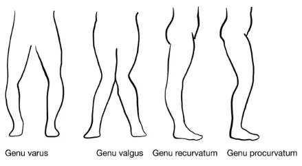
- Deformity:
- Genu Varum (Bow-Legged)
- Genu Valgum (Knock-Kneed)
- Genu Recurvatum (Hyperextension)
- Genu Procurvatum (Flexion deformity, always abnormal)
- Skin Changes and Scarring
Supine Position:
- Diffuse Swelling (e.g. knee effusion): Loss of parapatellar grooves/fossae; swelling to supra-patellar area
- Localized Swelling: Can indicate potential bursitis, tendon, or ligament pathology
Why Check: In a patient with chronic knee pain, the likelihood of osteoarthritis is greatly increased by bony enlargement (LR+ 11.8, LR- 0.5), and mildly increased by varus deformity (LR+ 3.4, LR- 0.8).
Palpation, including assessment for knee effusion
AFMC level Pre-clerkship 3 · Clerkship 4
- Commenting on: TESTCA – Tenderness, Effusions, Swelling, Temperature, Crepitus, & Atrophy
- Ottawa Knee Rule: Specifically palpate patella and fibular head for tenderness.
With Knee Extended (Right Knee shown in diagram):
- Temperature: Sweep with back of hand – Patella should be cooler than thigh above and tibia below.
- Patella Palpation: Feel undersides by tilting patella to each side, feel with fingers or thumb
- Palpating Anteriorly, From Superior to Inferior: Quadriceps most superior→ Quadriceps Tendon → Patella → Patellar Ligament → Tibial Tuberosity most inferior
- Palpating Popliteal Fossa Posteriorly: Check popliteal pulse and for swelling
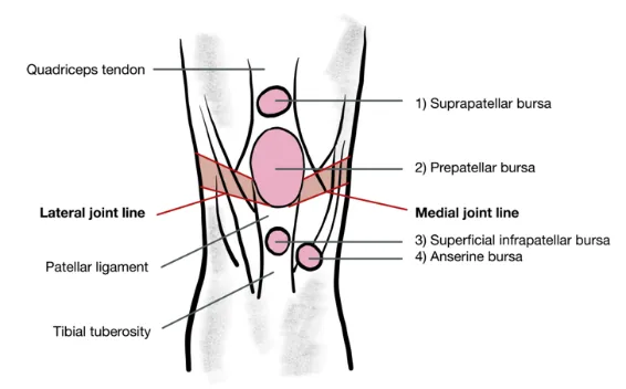
- Palpating 4 Bursae:
- Pre-Patellar Bursa (in front of patella)
- Superficial Infra-Patellar Bursa (inferior to patella, overlying patellar ligament and under skin)
- Deep Infra-Patellar Bursa (behind patellar ligament)
- Anserine Bursa (Anteromedial aspect of knee, near medial tibial plateau, deep to Sartorius, Gracilis and Semitendinosus Tendons)
With Knee Flexed to 90°:
- Palpate Joint Line:
- Medial & Lateral Joint Line; Medial Collateral Ligament (MCL)
- Medial & Lateral Tibial Plateaus; Femoral Condyles
- Adopt Figure of 4 Position (Heel resting on contralateral knee):
- Palpate Lateral Collateral Ligament (LCL), felt as cord on lateral knee aspect. Can feel hard as bone.
Palpation for Effusion:
- Fluid Wave Test or Bulge Sign (useful for smaller effusions): Sweep your hand up the medial aspect of the knee to empty the medial fossa. Then sweep down the lateral aspect. As you sweep down the lateral aspect, look for a bulge forming in the medial fossa. A bulge forming in the medial fossa indicates an effusion (may be false negative in larger effusions)
- For additional tests for Knee Effusions (Fluid Ballottement and Patellar Tap Tests) see Select Special Tests below
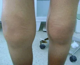
Bilateral Knee Effusion3
Why Check: Palpation can be very helpful to determine source of pain. Palpate for tenderness and swelling in a systematic manner, to assess for potential fractures and other abnormalities.
- Warm to touch: May indicate septic bursitis, inflammatory arthritis, or septic arthritis. Palpable increase in temperature significantly decreases likelihood of knee osteoarthritis (LR+ 0.3, LR- 1.6).
- Patellar tenderness: May indicate Patellofemoral Osteoarthritis or Patellofemoral Pain Syndrome.
- Swelling of Popliteal fossa: May indicate Baker’s Cyst
- Tenderness of Anserine Bursa: May indicate Pes Anserine Bursitis
- Tenderness of Medial and Lateral Joint Lines: May indicate Osteoarthritis or meniscal injury (LR+ 1.8, LR- 0.5)
- The likelihood of clinically significant knee fracture is increased by tenderness at head of fibula (LR+ 3.4), joint effusion (LR+ 2.5, LR- 0.5), or isolated patellar tenderness (LR+ 2.2, LR- 0.8).
- Both gout and pseudogout (CPPD) may present in the knee.
Range of Motion (ROM)
AFMC level Pre-clerkship 3 · Clerkship 4
- Approach: Perform Active ROM, and then Passive ROM if Active ROM is limited
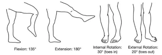
- Flexion (135°): Assess patellar crepitus during flexion-extension by placing 1 hand on patella
- Extension (180° – >10° hyper-extension past 180° is abnormal hypermobility)
- Internal Rotation (30°): Flex knee to 90°, point toes in
- External Rotation (20°): Flex knee to 90°, point toes out
- Patellar Movement: Push medially and laterally on patella; assess for hypermobility, pain or apprehension from dislocation.
- Patellar Compression test: With clinician hand just proximal to patella, press down and push patella distally, then ask patient to contract quadriceps – pain may indicate pain in patellofemoral compartment e.g. in patellofemoral osteoarthritis.
Why Check: Assessing passive range of motion if active range of motion is limited helps distinguish between ROM limitations caused by pain, tendon rupture etc. and those caused by structural limitation. Asymmetry with passive range of motion may indicate arthritis (bony end-feel), joint effusion (e.g. septic arthritis with soft end-feel), or internal derangements such as meniscal tear or fracture. The likelihood of clinically significant knee fracture is mildly increased by limited knee flexion <60º (LR+ 4.7, LR- 0.6) or limited knee flexion <90º (LR+ 2.9, LR- 0.5). The likelihood of meniscal injury is mildly increased by blocked full extension (LR+ 3.2, LR- 0.7), and pain on forced extension (LR+ 1.6, LR- 0.7).
Power Assessment
AFMC level Pre-clerkship 3 · Clerkship 4
- Approach: Resisted Isometric Testing i.e. patient resists force applied by clinician
- Sitting Approach: With knee flexed to 45-90° over side of exam table.
- Knee Extension: Examiner braces knee & pushes leg towards exam table, patient resists motion.
- Knee Flexion: Examiner braces knee & pulls leg away from exam table, patient resists motion.
- Note: Strength of internal and external tibial rotation usually not tested.
- Reporting: For each muscle group tested, assess per the Medical Research Council Power Grading. If it is all 5/5, you can say “Power was 5/5 bilaterally in both upper and lower limbs”, and report specific deficits. NOTE: This scale is detailed in Chapter 31 – Screening Neurological Examination
Special Tests:
Ligament Testing:
Anterior Drawer Test & Posterior Drawer Test
AFMC level Pre-clerkship 1 · Clerkship 3
Say: I will examine for ligament tears in the knee by applying pressure to the lower leg in 2 different directions. Please bend the knee to 90°, with the foot relaxed on the exam table.
Do:
- Patient supine, knee at 90° flexion. Stabilize tibia, grasping proximal tibia with thumbs on either side of joint line.
- Anterior Drawer: Apply anterior force to pull tibia forward.
- Posterior Drawer: Apply posterior force to push tibia backward. Look for excess displacement of tibia.
- Compare to other side.
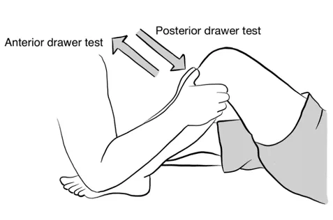
Report: No excess displacement of tibia seen – both anterior and posterior drawer tests are negative.
Why Check: A positive drawer test may increase the likelihood of anterior cruciate ligament (ACL) tear (LR+ 3.8-13.6, LR- 0.3-0.4). Some studies have shown that the LR+ may or may not be statistically significant. The Lachman Test (below) has consistently been found to have a higher LR+ for ACL tear across studies. A positive posterior drawer test greatly increases the likelihood of a suspected posterior cruciate ligament (PCL) tear (LR+ 97.8, LR- 0.1).
Lachman Test
AFMC level Pre-clerkship 1 · Clerkship 3
Say: I will continue to examine for ligament tears in the knee.
Do:
- Patient supine. Stabilize femur with one hand and grasp upper tibia with other hand with knee at 30 degrees flexion.
- Try to pull tibia forward. Assess for excess anterior movement of the tibia (or no discrete end point)
Report: No significant anterior movement of tibia seen – Lachman test is negative.
Why Check: A positive Lachman Test greatly increases the likelihood of a suspected anterior cruciate ligament (ACL) tear (LR+ 19.5-42, LR- 0.1-0.2). Studies have found it to consistently have a better LR+ and LR- than the anterior drawer test (above).
MCL Stress Test & LCL Stress Test
AFMC level Pre-clerkship 1 · Clerkship 3
Say: I will continue to examine for ligament tears in the knee.
Do: Patient supine. Knee at 0° initially and then 30° flexion, foot on table. Stabilize knee posteriorly with one hand. Stabilize lower leg with other hand.
- MCL stress test: Apply valgus force to lower leg with knee stabilized (push medially on knee, pull laterally on lower leg). Look for ↑ laxity and pain at MCL.
- LCL stress test: Apply a varus force to lower leg with knee stabilized (pull laterally on knee, push medially on lower leg). Look for ↑ laxity and pain at LCL.
Report: I note no increased laxity or pain in the MCL or LCL – both MCL and LCL stress tests are negative.
Why Check: A positive medial collateral stress test moderately increases the likelihood of a suspected medial collateral ligament tear (LR+ 7.7, LR- 0.2). A positive lateral collateral stress test greatly increases the likelihood of a suspected lateral collateral ligament tear (LR+ 16.2).
Meniscal Testing:
McMurray’s Test
AFMC level Pre-clerkship 1 · Clerkship 3
Say: I will examine for meniscal tears in the knee by supporting the leg and applying a pressure with the foot facing in and then facing out. Please keep the leg relaxed and let me know if you feel any pain during the test.
Do:
- Patient supine with leg relaxed. Clinician stands to side of patient to be tested (e.g. on the patient’s left side to test their left knee).
- With one hand, grasp the patient’s heel. Your hand grasping the patient’s heel will support the limb and provide required movement through the range of motion in McMurray’s test.
- With your other hand, grasp the patient’s knee, palpating the joint line with your thumb on one side of the joint line, and index finger on the other side of the joint line.
- To evaluate for a medial meniscus tear, move the patient’s hip and knee into flexion, then externally rotate and extend the knee.
- To evaluate for a lateral meniscus tear, move the patient’s hip and knee into flexion, then internally rotate and extend the knee
- These two motions can be done smoothly and in sequence, or each meniscus can be tested by separate discrete maneuvers. If pain is elicited, or a palpable or audible click occurs, this indicates a meniscal tear.
Report: I note no pain elicited or palpable or audible clicks – McMurray’s test is negative for a meniscal tear.
Why Check: A positive McMurray test mildly increases the likelihood of a suspected meniscal tear (LR+ 4.0, LR- 0.6), and has a Sensitivity of 71% and Specificity of 71% for meniscal tear. With a meniscal tear, patient will tend to localize pain either as a medial or lateral meniscus issue. You are trying to elicit pain and a popping or clicking sensation by trapping meniscus between femur and tibia.
- Medial Meniscus Tear: Pain with popping or clicking on external rotation and extending the knee
- Lateral Meniscus Tear: Pain with popping or clicking on internal rotation and extending the knee
- Note: Lateral meniscus is more mobile than medial meniscus → ↑ likelihood of lateral meniscus tear
- Osteoarthritis: Pain without popping or clicking during McMurray’s test can occur in Osteoarthritis
Apley’s Compression Test
AFMC level Pre-clerkship 1 · Clerkship 3
Say: I will continue to examine for meniscal tears in the knee by supporting the leg with the knee bent and applying a pressure with the foot facing in and then facing out. I will ask you to lie on the stomach for this test with the legs relaxed. Please let me know if you feel any pain during the test.
Do:
- Patient prone, knee flexed to 90 degrees, clinician stabilizes femur with one hand, grasps heel with other hand.
- Grasp patient’s heel and apply downward pressure through heel while rotating lower leg internally and externally. Apply a valgus force/tilt (medial) for medial meniscus, and a varus force/tilt (lateral) for lateral meniscus. If pain is elicited, or a popping/clicking noise occurs, this raises concern for a possible tear of the respective meniscus.
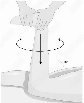
Report: I note no pain elicited or palpable or audible clicks – Apley’s compression test is negative.
Why Check: Apley’s test is a two-part test to assess medial or lateral knee pain. Apley’s Compression Test has a Sensitivity of 61% and Specificity of 70% for meniscal tear. With a meniscal tear, patient will tend to localize pain either as a medial or lateral meniscus issue. You are trying to elicit pain or “clunk” sensation by trapping meniscus between femur and tibia.
Apley’s Distraction Test
Say: I will examine for collateral ligaments tears in the knee pulling up on your lower leg while twisting the foot. I will ask you to lie on your stomach for this test with the legs relaxed. Please let me know if you feel any pain during the test.
Do:
- Patient prone, knee flexed to 90 degrees
- Clinician grasps the leg proximal to the ankle and applies traction to the tibia towards the ceiling, then applies internal and external rotation of the tibia while tractioning.
- If pain is elicited along either joint line, there may be a tear of the collateral ligament on that side
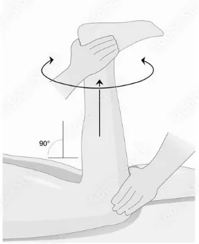
Report: I note no pain elicited – Apley’s distraction test is negative
Why check: Apley’s test is a two-part test to assess medial or lateral knee pain. Apley’s distraction test can help identify injury to the collateral ligaments.
Part 3: Select Special Tests – Not Part of Routine Knee Exam But Seen in Practice:
Additional Tests to Palpate for Knee Effusion
- Fluid Ballottement Test: With leg extended, grasp knee just above the patella with 1 hand. With other hand, grasp the medial and lateral parapatellar fossae. With hand just above patella, apply and release pressure, while feeling for ballotable fluid in other hand. If ballotable fluid, this indicates an effusion.
- Patellar Tap (useful for moderate-large effusions): Apply pressure with one hand just above the patella and sweep down until you reach the upper pole of the patella. This pushes fluid into the suprapatellar pouch behind patella. Keep pressure applied. With other hand, use 1-2 fingers to push the patella down, and assess whether it taps the femur and bounces back up. If patella taps the femur, then bounces back up, this indicates an effusion (may be false negative in smaller effusions)
Why Check: As mentioned above, the Fluid Wave Test or Bulge Sign is most useful for smaller effusions and routinely used in clinical practice. Two additional approaches to palpation for knee effusions are the Fluid Ballottement and Patellar Tap tests.
Pivot Shift Test
Say: I will continue to examine for ligament tears in the knee.
Do:
- Patient supine with hips and knees extended, relaxed. Brace patient’s ankle while keeping knee in extension. Internally rotate ankle/foot to internally rotate tibia and apply a valgus force (push medially on the fibula).
- Slowly flex the patient’s knee; a positive test occurs if tibia subluxes posteriorly (a pivot shift) which may be seen as anterior knee reducing or felt as a clunk (typically 45 degrees flexion).
Report: The pivot shift test is negative for ACL Tear.
Why Check: The Pivot Shift Test is used to assess for ACL Tears. To be diagnostic, it requires the patient to have an intact Iliotibial Band and MCL, and ability to extend knee to almost full extension (e.g. difficult to perform in presence of comorbid meniscal tear). A positive pivot shift test moderately increases the likelihood of suspected ACL tear (LR+ 8.8, LR- 0.7).
Clarke Test (Or Patellofemoral Grinding Test)
Say: I will examine the knee for a functional pain called Patellofemoral Pain Syndrome (PFPS).
Do:
- Patient supine, knees supported by pad to create 10-20 degrees of knee flexion, articulating patella in patellofemoral joint. Clinician presses on superior border of patella, applying force distally.
- Ask patient to contract quadriceps muscle. Ask the patient whether pain is reproduced during test.
Report: Clarke’s Test is negative as it did not reproduce the patient’s pain.
Why Check: The Clarke Test is used to assess for Patellofemoral Pain Syndrome (PFPS). A positive Clarke test mildly increases the likelihood of suspected PFPS (LR+ 1.18, LR- 0.7-0.91).
Patellar Apprehension/Displacement Test
Say: I will continue to examine the knee for a functional pain called Patellofemoral Pain Syndrome (PFPS).
Do:
- Patient supine, relaxed with knee in full extension. Clinician presses on medial patella to displace laterally for a lateral patellar guide, grasping ankle on same side with other hand.
- Perform slow combined flexion of knee and hip, keeping lateral pressure applied on patella for lateral patellar tracking. Ask patient whether their pain is reproduced and watch for signs of apprehension.
Report: Patellar Apprehension Test is negative; it did not cause apprehension or reproduce patient’s pain.
Why Check: Assess for PFPS. An alternative method is Patellar Displacement Test – patella displaced medially to assess pain improving through passive ROM. The evidence for Patellar Apprehension test suggests it is not clinically significant for detecting PFPS (LR+ 0.9-2.3, LR- 0.8-1.0).
Patellar Tilt Test
Say: I will continue to examine the knee for a functional pain called Patellofemoral Pain Syndrome (PFPS).
Do:
- Patient supine, relaxed with knee in full extension. Observe for resting tilt of patella with medial side more anterior than lateral side. Clinician applies pressure on anteromedial patellar border in posterior direction, and also applies pressure on lateral patellar border in anterior direction.
- Assess whether patella corrects tilt to neutral at minimum (Normal tilt 15 degrees medio-laterally).
Report: The patient has a normal tilt on the Patellar Tilt Test.
Why Check: The Patellar Tilt Test is used to assess for Patellofemoral Pain Syndrome (PFPS). If tilt does not correct with pressure to neutral position, this indicates excessive lateral tightness. A positive patellar tilt test moderately increases the likelihood of suspected PFPS (LR+ 5.4, LR- 0.6).
Functional Trendelenburg Test
Say: I will examine the hip for weak hip abductors, which can cause anterior knee pain.
Do:
- Patient stands on affected leg in single leg stance; can hold onto an item for support if balance concern.
- Patient does a shallow “squat” with knee flexed to 20 to 30 degrees. If knee moves into valgus, the test is positive. You may also see the contralateral pelvis drop (as seen in Trendelenburg Gait)
Report: The patient was able to flex their knee in a single-leg stance without the knee moving into Valgus – the Functional Trendelenburg test is negative.
Why Check: The Functional Trendelenburg Test is typically used to assess for weakness in the hip abductor muscles (Gluteus Medius and Gluteus Minimus), which can contribute in a kinetic chain to anterior knee pain such as Patellofemoral Pain Syndrome (PFPS). It can also occur with congenital hip dislocation, as well as arthritis (both rheumatoid and osteo-arthritis).
Helpful Videos to Review:
- Video 1: Knee Exam – McMaster MSK Examination Series (Link: KNEE EXAMINATION - McMASTER UNIVERSITY)
- Video 2 and 3: Bates’ Visual Guide to the Physical Exam – Anatomy Review and Examining the Knees
- Video 4 and 5: Dr. Janice Harvey’s Joint Assessment Made Easy – Knee Assessment Part 1 and 2
Part 4: Relevant Likelihood Ratios – from Rational Clinical Examination JAMA series
Clinical Question 1: Does this patient have a Meniscal Tear?4
| Procedure | Sensitivity | Specificity |
|---|---|---|
| McMurray’s Test | 0.71 | 0.71 |
| Apley’s Test | 0.61 | 0.702 |
Clinical Question 2: Does this patient have an ACL Tear?5
| Test (n = No. of studies) | LR+ (95% CI) | LR- (95% CI) |
|---|---|---|
| Lachman Test (n=1) | 42 (2.7-651) (about +45%) | 0.1 (0.0-0.4) (about −45%) |
| Anterior Drawer Test (n=3) | 3.8 (0.65-22) (no change: 95% CI includes 1) | 0.3 (0.05-1.5) (no change: 95% CI includes 1) |
Clinical Question 3: Does this patient with Chronic Knee Pain have Osteoarthritis?6
| Procedure | LR+ (95% CI) | LR- (95% CI) |
|---|---|---|
| Bony enlargement | 11.8 (4.9-28.2) (about +45%) | 0.5 (0.4-0.6) (about −15%) |
| Varus deformity | 3.4 (1.6-7.6) (about +15%) | 0.8 (0.7-0.9) (about −15%) |
| Stiffness <30 minutes | 3.0 (2.1-4.4) (about +15%) | 0.2 (0.1-0.3) (about −30%) |
| Crepitus, passive motion | 2.1 (1.7-2.7) (about +15%) | 0.2 (0.1-0.3) (about −30%) |
| Valgus deformity | 1.4 (0.8-2.4) (no change: 95% CI includes 1) | 0.9 (0.8-1.0) (no change: 95% CI includes 1) |
| Palpable increase in temperature | 0.3 (0.2-0.5) (about −30%) | 1.6 (1.4-2.0) (about +15%) |
| At least 3 of 6 findings above | 3.1 (2.3-4.1) (about +15%) | 0.1 (0-0.1) (about −45%) |
Clinical Question 4: Does this patient have a Clinically Significant Knee Fracture?6
*Clinically Significant: Requiring ortho consult, splinting, surgery (>5mm fracture or assoc. complete tendon, ligament disruption)
| Procedure | LR+ (95% CI) | LR- (95% CI) |
|---|---|---|
| Limitation of knee flexion <60 degrees | 4.7 (3.8-5.9) (about +15%) | 0.6 (0.5-0.7) (about −15%) |
| Inability to bear weight, immediately and in ER | 3.6 (3.0-4.3) (about +15%) | 0.6 (0.5-0.7) (about −15%) |
| Tenderness at head of fibula | 3.4 (2.5-4.7) (about +15%) | 0.9 (0.8-1.0) (no change: 95% CI includes 1) |
| Age >55 years’ old | 3.0 (1.6-5.3) (about +15%) | 0.7 (0.5-1.0) (no change: 95% CI includes 1) |
| Limitation of knee flexion <90 degrees | 2.9 (2.5-3.4) (about +15%) | 0.5 (0.4-0.7) (about −15%) |
| Joint effusion | 2.5 (2.0-3.0) (about +15%) | 0.5 (0.3-0.7) (about −15%) |
| Isolated tenderness of patella | 2.2 (1.6-2.9) (about +15%) | 0.8 (0.8-0.9) (about −15%) |
| Ecchymosis | 2.2 (0.9-5.3) (no change: 95% CI includes 1) | 0.9 (0.7-1.1) (no change: 95% CI includes 1) |
| Ottawa Rule positive (See below) | 1.7 (1.4-2.1) (about +15%) | 0.1 (0-0.3) (about −45%) |
Clinical Question 5: Does This Patient have Patellofemoral Pain Syndrome (PFPS)?7
| Procedure | Sensitivity | Specificity | LR+ | LR- |
|---|---|---|---|---|
| Patellar Tilt Test | 0.43 | 0.92 | 5.4 (about +30%) | 0.6 (about −15%) |
| Pain During Squatting | 0.91 | 0.50 | 1.8 (about +15%) | 0.2 (about −30%) |
| Clarke Test | 0.39-0.48 | 0.67-0.75 | 1.18 (about +15%) | 0.7-0.91 (about −15%) |
| Patellar Apprehension Test | 0.07-0.32 | 0.86-0.92 | 0.9-2.3 (about −15%) | 0.8-1.0 (about −15%) |
| Medial or Lateral Patellar Facet Tenderness | 0.92 | 0.65 | N/A | 0.96 (about −15%) |
Clinical Question 6: Does This Patient have a Ligamentous or Meniscal Knee Injury?8
| Procedure | Sensitivity | Specificity | LR+ | LR- |
|---|---|---|---|---|
| ACL: Lachman Sign | 0.48-0.96 | 0.90-0.99 | 19.5 (about +45%) | 0.2 (about −30%) |
| ACL: Anterior Drawer Sign | 0.27-0.94 | 0.91-0.99 | 13.6 (about +45%) | 0.4 (about −30%) |
| ACL: Pivot Shift Test | 0.06-0.61 | 0.95-0.99 | 8.8 (about +30%) | 0.7 (about −15%) |
| PCL: Posterior Drawer Sign | 0.90-0.95 | 0.99 | 97.8 (about +45%) | 0.1 (about −45%) |
| Meniscal: McMurray Sign | 0.17-0.80 | 0.77-0.98 | 4.0 (about +15%) | 0.6 (about −15%) |
| Meniscal: Block to full extension | 0.44 | 0.86 | 3.2 (about +15%) | 0.7 (about −15%) |
| Meniscal: Joint Line Tenderness | 0.55-0.92 | 0.30-0.83 | 1.8 (about +15%) | 0.5 (about −15%) |
| Meniscal: Pain on forced extension | 0.47-0.51 | 0.67-0.70 | 1.6 (about +15%) | 0.7 (about −15%) |
| MCL: Valgus stress laxity | 0.79-0.89 | 0.49-0.99 | 7.7 (about +30%) | 0.2 (about −30%) |
| LCL: Varus stress laxity | 0.25 | 0.98 | 16.2 (about +45%) | Not significant |
References
-
Ottawa Knee Rule. MDCalc. Accessed March 12, 2022. https://www.mdcalc.com/ottawa-knee-rule#next-steps ↩ ↩2
-
Recommendations. Choosing Wisely Canada. Accessed July 22, 2022. https://choosingwiselycanada.org/recommendations/#list ↩ ↩2
-
Hegedus EJ, et al. Physical examination tests for assessing a torn meniscus in the knee: a systematic review with meta-analysis. J Orthop Sports Phys Ther. 2007;37(9):541-550. ↩
-
Does this patient have an ACL tear – update. Rational Clinical Examination Series, chap 28. 2009:370. ↩
-
McGee S. Evidence Based Physical Diagnosis. Saunders; 2012:834-5. ↩
-
Gaitonde D, Ericksen A, Robbins R. Patellofemoral pain syndrome. Am Fam Physician. 2019;99(2):88-94. ↩ ↩2
-
McGee S. Evidence-Based Physical Diagnosis. 4th ed. Elsevier; 2012:chap 64. ↩