Integration Foundation
Chapter 39Pre-Operative Anesthesia Assessment
Introduction
- Key Idea – 4 As of Anesthesia: Controlled and reversible states of
- Awareness (Lack of): Unconsciousness
- Amnesia: Lack of memory of the event
- Analgesia: Abolition of subconscious reactions to pain (including somatic reflexes such as moving or withdrawing, and autonomic reflexes such as hypertension, tachycardia, sweating)
- Akinesia: Lack of overt movement; muscle relaxation may be required in some cases
- Pre-Operative Anesthesia Assessments are important for identifying risk factors and appropriate planning to manage risks posed by the proposed surgical intervention. This visit is aimed to initiate the doctor-patient relationship; understand the patient’s History of Presenting Illness and Past Medical History; review medications; discuss personal and family anesthetic history including any issues; identify potential intraoperative problems (e.g. airway management issues); order investigations and optimize patient; allay fears and present a patient-centred plan; and plan post-op disposition.
- For more info about Anesthesia for Medical Students, helpful resources are:
- Raymer, K. 2012. Understanding Anesthesia: A Learner’s Guide [Internet]. (Available Free on Medportal)
- Sullivan, P. 2012. The Ottawa Anesthesia Primer. Echo Book Publishing (Available for Purchase Online)
- Resource for Disease-Specific Anesthesia Considerations: https://www.anesthesiaconsiderations.com/
- New York School of Regional Anesthesia (NYSORA): https://www.nysora.com
Part 1: Elective Surgery – Approach to Pre-Op Assessments
Patient Selection – Identifying Patients who benefit from a Pre-Operative Clinic Visit
Key Idea: Not all patients need a pre-op clinic visit. The following algorithm, adapted from the Choosing Wisely 2019 Drop the Pre-Op Toolkit1, is helpful in identifying patients who would benefit from a Pre-Op visit based on planned surgical procedure (e.g. invasiveness), and patient’s physiological status (e.g. ASA Classification).
Step 1 – What is the Physiological Status of the Patient?
The American Society of Anesthesiologists (ASA) Classification is a risk-stratification tool assessing patient pre-op status to help plan surgical interventions, and to communicate overall physical condition of patient, as follows:
| ASA | Description | Examples |
|---|---|---|
| I | Healthy patient | No organic, physiological, biochemical or psychiatric disturbance |
| II | Mild systemic disease – no functional limitation | Heart disease slightly limiting physical activity; hypertension; diabetes; chronic bronchitis; anemia |
| III | Severe systemic disease – definite functional limitation | Heart or chronic pulmonary disease limiting activity; poorly controlled hypertension; diabetes on insulin ± complications; angina pectoris; history of MI; OSA ± CPAP; cancer |
| IV | Severe systemic disease – a constant threat to life | CHF; persistent angina pectoris; advanced pulmonary; renal or hepatic dysfunction; recent TIA |
| V | Moribund patient – not expected to survive 24hr without operation | Massive trauma; ruptured AAA; cerebral hemorrhage with mass effect. |
| VI | Declared brain-dead patient, organs being removed for donor purposes | - |
| E | Added suffix to ASA Category to denote Emergency procedure | - |
In addition, ask – Does this patient have unique perioperative care considerations? For example:
- Anticoagulant medications ± bridging required
- History of problems with anesthetics
- Obstetrical patient with comorbidity
- Coagulopathy
- Polypharmacy
- Jehovah’s Witness
- Airway concerns
- Chronic pain
- Planned ICU admission
Finally, ask – Would this patient benefit from a Medicine Consult? For example:
- Coronary Artery Disease (CAD)
- Use of Antiplatelet (e.g. Aspirin, P2Y12 Inhibitors) or Anticoagulant (Warfarin, DOAC) Medications
- Need for Endocarditis Prophylaxis (GI/GU/Oral Procedure with Hx of Inf. Endocarditis, Prosthetic Valve, Cyanotic Heart Disease, Heart Transplant)
- ↑ Cardiac Risk (≥ 2 of: CAD, CHF, CKD, DM on Insulin, TIA/Stroke)
- ↑ Respiratory Risk (Asthma/COPD with recent/frequent exacerbations, ILD)
- Use of Steroids (or recent taper) or immunosuppressive meds
- Use of Insulin or >2 Oral Agents for Type 1/Type 2 Diabetes
Step 2 – What is the Surgical Intervention Proposed?
More invasive surgeries carry ↑ risk of significant blood loss, cardiorespiratory compromise, ↑ time, unusual patient positioning, etc. and benefit from Pre-Op Visits. A scheme categorizing surgeries based on invasiveness with examples is as follows:
ENT
- Minimally Invasive – Myringotomy, Microlaryngoscopy, Bronchoscopy
- Min-Mod Invasive – T&A, Mastoid, Septo/Rhinoplasty, Sinus
- Mod-Significantly Invasive – Thyroidectomy, Parotidectomy
GEN
- Minimally Invasive – Port Insertion
- Min-Mod Invasive – Ano-Rectal procedure, Hernia, Lap Chole/Appendectomy/Hernias/Lysis, Breast Biopsy/ Needle Loc, Mastectomy
- Mod-Significantly Invasive – Open Chole, Major Lap surgery (stomach/spleen/bowel), Open bowel resection
- Highly Invasive – Major bowel resection, Major VATS or open thoracic, esophagectomy
GYNE
- Minimally Invasive – Hysteroscopy
- Min-Mod Invasive – Dilation and Curettage (D&C), Laparoscopy
- Mod-Significantly Invasive – Hysterectomy/Myomectomy, Vaginal Hysterectomy/repair
OPTHO
- Minimally Invasive – Lens surgery with local anesthetic and sedation
ORAL
- Min-Mod Invasive – Dental/Restorations Extractions/biopsy
- Mod-Significantly Invasive – Oro/Maxillofacial
ORTHO
- Min-Mod Invasive – Arthroscopy (except shoulder)
- Mod-Significantly Invasive – Extremities
- Highly Invasive – IM Nailing, Hip & Long Bone Fractures, Amputations
- Always requiring Pre-Op Visit – Arthroplasty (Hip/Knee/Shoulder); Shoulder arthroscopy
PLASTICS
- Minimally Invasive – Extremities
- Mod-Significantly Invasive – Breast reduction
PSYCH
- Minimally Invasive – ECT
UROL
- Minimally Invasive – Circumcision, Vasectomy, Cystoscopy
- Mod-Significantly Invasive – Nephrectomy, TURP
- Highly Invasive – Radical Prostate
VASC
- Min-Mod Invasive – Vein Ligation/Stripping
- Highly Invasive – Bypass, Aneurysm Repair, Endarterectomy
Step 3 – Decide who would benefit from a Pre-Op Visit
Based on Steps above, the Choosing Wisely 2019 Drop the Pre-Op Toolkit1 recommends triaging cases as follows:
- Category 1 – No Pre-Op Visit
- Category 2 – Pre-Op Visit with Registered Nurse (RN) Only
- Category 3 – Pre-Op Visit with RN, Anesthesia ± Medicine (re. Would this patient benefit from a Medicine Consult?)
| Min Invasive | Min-Mod Invasive | Mod-Sign. Invasive | Highly Invasive | |
|---|---|---|---|---|
| ASA I | Category 1 | Category 1 | Category 2 | Category 3 |
| ASA II | Category 1 | Category 2 | Category 2 | Category 3 |
| ASA III | Category 3 | Category 3 | Category 3 | Category 3 |
| ASA IV | Category 3 | Category 3 | Category 3 | Category 3 |
| Unique Pre-Op Care Required | Category 3 | Category 3 | Category 3 | Category 3 |
Focused History – Based on Chief Complaint (Begin with Open-Ended Questions)
Anesthesia History
- Any previous history of general anesthetic?
- If yes, any previous problems with general anesthesia (e.g. Airway management issues, post-op nausea/vomiting, malignant hyperthermia, pseudocholinesterase deficiency)?
- Ensure you have up to date consent for blood products.
Difficult Intubation [1, 2] or ventilation [2, 3]
- History of difficult intubation or bag-mask ventilation?
- Snoring or obstructive sleep apnea? (Screen with STOP-BANG (below))?
- C-spine (neck) surgery, pain, stiffness, arthritis or limited mobility?
- Dental alterations (caps, crowns, bridges, dentures)?
Exercise Tolerance
- Quick Function Screen for Functional Limitation: Can you climb 1 flight of stairs without stopping or becoming short of breath? (Metabolic Equivalency of ADL – 5)
- 3 important sets of exertional symptoms to screen for:
- Chest Pain (exertion or at rest + associated dyspnea, N/V, palpitations, presyncope, syncope)
- Heart Failure (orthopnea, PND, peripheral edema)
- Dyspnea (SOB at rest or exertion)
CCS (Canadian Cardiovascular Society) Functional Classification of Angina
| CCS Class | Description |
|---|---|
| I | Ordinary physical activity does not cause angina (e.g. walking, climbing stairs). Angina with strenuous, rapid, prolonged exertion. |
| II | Slight limitation of ordinary physical activity. Angina walking >2 blocks on level surface, climbing > 1 flight of ordinary stairs (normal pace, conditions). |
| III | Marked limitation of ordinary physical activity. Angina walking ≤2 blocks on level surface, climbing ≤ 1 flight of ordinary stairs (normal pace, conditions). |
| IV | Inability to carry on any physical activity without discomfort. Angina may be present at rest. |
NYHA (New York Heart Association) Functional Classification of Heart Failure
| NYHA Class | Description |
|---|---|
| I | Ordinary physical activity does not cause heart failure symptoms |
| II | Comfortable at rest; ordinary activity results in heart failure symptoms |
| III | Market limitation of ordinary activity; less than ordinary activity results in heart failure symptoms |
| IV | Inability to carry on any physical activity without discomfort. Symptoms may be present at rest. |
MRC (Medical Research Council) Breathlessness Scale for Dyspnea
| MRC Grade | Description |
|---|---|
| 1 | Not troubled by breathlessness except with strenuous exercise |
| 2 | Troubled by breathlessness when hurrying on level, walking up slight hill |
| 3 | Walks slower than people of same age on level due to breathlessness, or has to stop for breath when walking at own place on the level |
| 4 | Stops for breath after walk ~90 meters or after few minutes on level |
| 5 | Too breathless to leave house, or breathless when dressing/undressing |
Obstructive Sleep Apnea (OSA) Screen – STOP-BANG Questionnaire
– See Ch 7 – Head & Neck Exam – MF1
- Snoring: Do you snore loudly (loud enough to be heard through closed doors or your bed-partner elbows you for snoring at night)?
- Tired: Do you often feel tired, fatigued or sleepy during the daytime (such as falling asleep during driving or talking to someone)?
- Observed: Has anyone observed you stop breathing or choking/gasping during sleep? Pressure: Do you have or are being treated for high blood pressure?
- Pressure: History of/Treatment for Hypertension?; BMI: >35kg/m²?; Age: >50yo?
- Neck Size Large (Measured around Adams Apple): For males, is your shirt color 17”/43cm or larger? For females, is your shirt collar 16”/41cm or larger?
- Gender: Male?
Time Since Last Meal3
- Recommendations to ↓ Perioperative Aspiration Risk Day of:
- Clear Liquids (Water, juice no pulp, carbonated drinks, black coffee, clear tea): 2 hours
- Breast Milk: 4 hours vs. Infant Formula: 6 hours
- Nonhuman Milk: 6 hours
- Light Meal (Toast and clear liquids): 6 hours
- Heavy Meal or other foods: 8+hours may be needed
Medical Profile
For each chronic illness – assess severity, stability, complications, functional limitation
Cardiovascular
- Coronary Artery Disease (CAD) – recent MI, unstable angina? History of Coronary Artery Bypass Graft, Coronary Artery Stents?
- Other Conditions to Assess4: Peripheral Artery Disease (PAD), Heart Failure (HF) or Severe Obstructive Intracardiac Abnormality (Aortic Stenosis, Mitral Stenosis, HOCM); Arrhythmias (e.g. Atrial Fibrillation – Anticoagulation re. CHADSVASC risk, HAS-BLED Score? Rate vs. rhythm control?)
- Devices: Permanent Pacemaker (PPM); Implantable Cardioverter-Defibrillator (ICD); Left Ventricular Assist Device (LVAD)?
Hematology
- Conditions to Assess: Anemia; Coagulopathies; Blood dyscrasias
Respiratory
- COPD5 – Using GOLD Classification – ask about symptoms/exercise capacity with MRC scale (see below – MRC Grade ≤2 = less symptomatic; MRC Grade ≥ 3 = more symptomatic); COPD Exacerbations in past year (Classic triad: ↑ breathlessness, ↑ sputum production, ↑ sputum purulence) including hospitalizations; medication adherence; last pulmonary function tests (PFTs), Arterial Blood Gases (re. whether patient is a CO2-retainer); on Home O2 therapy?; and smoking
- Asthma6: Assess Control (Well-Controlled if – No daytime asthma symptoms, no nighttime asthma symptoms, no physical activity restriction due to asthma, no missed school/work due to asthma, no need for reliever unless prescribed for exercise); Triggers; Medication Adherence; last PFTs performed; and Exacerbations (previous ER visits, hospitalizations, ICU admissions, Intubations for asthma exacerbations). What inhalers do you use? When did you last take them?
- Sleep Apnea7: Assess lifestyle measures (weight loss, sleep position, alcohol use); CPAP or BiPAP (adherence, date of last CPAP/BiPAP titration); Mandibular Advancement Devices (adherence, date of last fit); Epworth Sleepiness Scale (re. severity of daytime sleepiness); concerns with driving safety
- Other Conditions to Assess: Interstitial Lung Disease; Pulmonary Hypertension (PHTN)
Renal
- General: CKD Stage? Most recent Creatinine Clearance? Lifestyle modifications? Current medications renally dosed?
- Dialysis: Hemodialysis (HD – time HD day prior to surgery) vs. Peritoneal Dialysis (PD)?
- Complications: Hypertension; Anemia; ↑ PTH; ↑ Ca; ↑ K; ↑ PO4; Metabolic Acidosis; Heart Failure/Cardio-Renal Syndrome; LV Hypertrophy
Gastrointestinal
- Diseases increasing risk of perioperative aspiration pneumonitis8: GI Obstruction; Delayed gastric emptying (e.g. gastroparesis, previous GI surgery, opioids, recent trauma, pregnancy); Incompetent Lower Esophageal Sphincter (e.g. GERD, dyspepsia, hiatal hernia, previous upper GI surgery, pregnancy); Morbid Obesity
- Hepatic Disease – potential to impact intraoperative drug metabolism, coagulation status, fluid status. (e.g. Cirrhosis – Etiology; Hx Decompensating events e.g. Ascites, Spontaneous Bacterial Peritonitis, Esophageal Varices, Hepatic Encephalopathy, Hepatorenal Syndrome; Recent MELD Score i.e. 3-month mortality risk)
Endocrine
- Diabetes – Age of Dx; Glycemic Control (Hx DKA, HHS? A1c, FBG; CBG Checks at home/results?); Management (Insulin? Oral Agents?); Microvascular (neuropathy, nephropathy, retinopathy) and Macrovascular Complications (CAD, CVD, PVD)
- Thyroid: Hypo/Hyperthyroidism (Date of last TSH? Symptoms? Medication doses?); Goiter (Mass effect symptoms)
- Pheochromocytoma (Classic Triad: Headache, Palpitations, Diaphoresis) – Risk of labile BP and HR throughout perioperative period, requiring careful preoperative adrenergic blockade management.
- Hyper/Hypocortisolism – Endogenous cortisol production ↑ in response to perioperative stress, can be blunted by history of exogenous corticosteroid use, i.e. ↑ risk of perioperative adrenal insufficiency.
Pregnancy
- Any chance the patient could be pregnant? (Consider Urine β-HcG if appropriate)
MSK
- Conditions to assess for: Conditions that can impact intubation e.g. Inflammatory Arthritis (e.g. Rheumatoid Arthritis); C-Spine Trauma; Trisomy 21 (including Atlantoaxial instability, Macroglossia, Subglottic Stenosis, Cardiovascular anomalies including septal defects)9; Myopathies; Dystrophies
Neurology
- TIA and CVD – Frequency of events; persistent sensory/motor deficits; if recent TIA – ABCD² Score for 2-day, 7-day, and 90-day stroke risk
- Other Conditions to Assess: Seizures (Type, frequency, date of last seizure); Spinal Cord Injury (Date, Level of Neurologic injury); Neuromuscular Junction (NMJ) Diseases (e.g. Myasthenia Gravis); ↑ Intracranial Pressure (ICP)
Family Medical History (FMHx)
Particularly important if patient has no history of exposure to general anesthesia
- History of Malignant Hyperthermia: Rare, heritable clinical syndrome in response to Succinylcholine (depolarizing muscle relaxant) or Inhaled Anesthetics (exception being Nitrous Oxide).
- History of Pseudo-cholinesterase Deficiency: A heritable deficiency in the enzyme pseudocholinesterase, which breaks down two neuromuscular blocking agents: succinylcholine and mivacurium. This heritable enzyme deficiency causes prolonged paralysis when using succinylcholine or mivacurium.
Social Hx
Smoking
Pack years = packs/day*yrs…Have you thought about quitting within the next 6 months? How long ago did you quit smoking? 5As approach taking a smoking history
- Pre-Operative encounter is an opportunity for smoking cessation: Discuss and facilitate smoking cessation (e.g. Nicotine replacement) ideally ≥ 4 weeks before surgery1
- Longer duration of smoking cessation is more effective in reducing postoperative complications10
- Day of surgery, ask: When was your last cigarette?
Alcohol
How Often Do You Drink (Drinks/Week)? What is the size of 1 drink? What kinds of drinks?
- Red Flag Levels: 10 drinks/week for women, 15 drinks/week for men
- CAGE Screening Tool – 2 or more positive = investigate for alcoholism: Ever felt like you should cut down? Are you annoyed when someone criticizes you? Do you feel guilty? Do you need an eye opener in the morning?
- History of Alcohol Withdrawal, Seizures, Korsakoff’s/Wernicke’s– ↑ perioperative risk
- CIWA-Ar – Objectively quantifies alcohol withdrawal severity
- Most recent drink? What symptoms do you feel when you don’t drink for a while? If pt is at high risk of withdrawal, may counsel to drink night prior to surgery and focus on cessation afterwards.
Recreational Drugs
(See Ch 37 – Substance Use Disorders – MF4 for more expanded substance use Hx)
List off to normalize…Do you now use, or have you used any of the following substances:
- Marijuana (grams dried cannabis per day/week, # joints per day/week) - Red Flag: 3 joints/week
- Prescription drugs for non-medical purposes
- Street drugs (e.g. cocaine, ecstasy, heroin, MDMA, etc.)
- Stages of Change (Motivational Interviewing): Pre-contemplation, Contemplation, Determination, Action, Maintenance
Supports and Screening for Equity-Deserving Populations
Consider impact of social determinants of health (SDoH) on lived experiences of patients and their ability to implement preoperative recommendations and to access surgical care. You can help advocate for your patient by screening for SDoH and by learning about local resources. Using the IF-IT-HELPS Social History Tool11, additional questions include but not limited to:
- Identity – Tell me about yourself
- Family and Friends – Are there people in your life you can count on for support?
- Income – Do you ever have difficulty making ends meet at the end of the month?
- Housing Instability – Where are you staying right now? What is your housing like?
- Food Insecurity – Do you ever run out of money to buy food?
- Transportation – How are you managing getting to and from our appointments together? Do you have trouble paying for public transportation?
- Health Benefits – Do you have health coverage and benefits e.g. drug, physio coverage?
- Employment – Are you working right now?
- Education – How far did you go in your education? Do you have any trouble reading/writing?
Diet and Exercise
- How is your nutrition? What do you do to keep active? Compliment exercise if expressed…
Medications Hx, Allergies
In general, medications continued until day of surgery. Specific examples listed below.
Focused Allergy History
- Allergies to Anesthetics (e.g. Muscle Relaxants) & Latex Allergy
- If endorses allergy, qualify the type of reaction (e.g. hives vs anaphylaxis).
Endocrine
- Insulin – Generally give preoperative fasting details, insulin orders e.g. half AM insulin dose
- Oral Antihyperglycemics: Generally held on day of surgery
Neurology
- Lithium (Choosing Wisely1) – Check Lytes & Creatinine pre-operatively
Cardiovascular
- Aspirin (Thrombosis Canada12): Held 5-7 days pre-op for non-urgent non-cardiac surgery, restart 8-10 days post-op once surgical bleeding risk ↓
- Exception: patients with coronary stents undergoing elective/non-urgent noncardiac surgery: ASA continued perioperatively. Bare metal stent: delay surgery for 1mo after PCI. Drug eluting stent: delay surgery at least 3mo after PCI.
- Clopidogrel, Ticagrelor (Thrombosis Canada12– e.g. patients on Dual Antiplatelet Therapy): Held 5-7 days pre-op, restart once safe as per surgeon.
- DOACs (Thrombosis Canada13): For Low Bleeding Risk Surgery – likely safe not to interrupt DOAC, if concerned can delay dose that day to 4-6 hours post-procedure.
| Agent | Low/Mod. Bleed Risk Surgery | High Bleed Risk/Neuraxial |
|---|---|---|
| Pre-Op: Rivaroxaban, Apixaban, Edoxaban | Hold 2 days pre-op | Hold 3 days pre-op |
| Pre-Op: Dabigatran | CrCl ≥ 50: Hold 2 days pre-op CrCl 30-49: Hold 3 days pre-op | CrCl ≥ 50: Hold 3 days pre-op CrCl 30-49: Hold 5 days pre-op |
| Post-Op (All) | Start 24hrs post-op | Start 2-3 days post-op, consider interim prophylactic dose anticoagulants |
- Warfarin: See Thrombosis Canada Guidelines14 including risk-stratification; INR check day before surgery; bridging with Heparin (LMWH SC / UFH IV)
- β-Blocker (CCS4) – Generally continued; if low systolic BP, ↓ dose/withhold pre-op
- ACEi/ARB (CCS4) – Withheld 24 hours pre-op, restart post-op day 2 (if stable)
- Statin (CCS4) – Generally continued
- Digoxin, Diuretics (Choosing Wisely1) – Check Lytes & Creatinine pre-operatively
Important Note – Goals of Care in Pre-Operative Assessment
- We will explore general tools for holding a Values-Based goals of care conversation with the patient in Chapter 41 – Palliative Care Assessment. Unique elements to consider in preoperative goals of care discussions include the patient’s understanding of the surgery in addition to their underlying illness, and shared understanding of possible range of surgical outcomes15.
- A patient may have a Do-Not-Resuscitate (DNR) order documented, where surgery is offered and is within the patient’s goals of care. These scenarios are challenging but important discussions to have with patients and loved ones. It is worth discussing how such conversations are held while on your rotation. “The perioperative period is unique due to its acuity, the significant risks of surgery and anesthesia, potential for rapid treatment escalation, and the requirement for timely informed consent. It presents an opportunity for clinicians to engage patients in an honest discussion about their goals of care and wishes for their health.”16
Part 2: Pertinent Notes about the Physical Exam
- Draping: Drape appropriately when need area exposed; return covered up state at end of portion of exam/end of exam.
Risk factors of difficult intubation (LEMON)
- Look: Externally, chin, dentition, facial trauma, mouth opening
- Evaluate: 3-3-2 rule and mandibular protrusion
- Mallampati: Higher class increases risk
- Obstructions: May interfere with endotracheal tube
- Neck Mobility: C-spine surgeries, arthritis, stiffness, etc. may make it difficult to obtain sniffing position for intubation.
- AFMC Expectation Level: [1, 2]
Risk factors of difficult bag-mask ventilation (BONES)
- Think: Santa Claus is difficult to Bag-Mask Ventilate
- Beard: Can interfere with seal of mask with patient’s face
- Overweight/Obesity: Risk of upper airway obstruction from oropharyngeal tissue
- No Teeth: Prevents collapse of tongue against palate when mouth closed, and may be difficult to obtain seal
- Elderly (Age > 55yo): Decreased tissue elasticity, ↑ incidence of lung diseases decreasing lung compliance (COPD, Asthma, Pulmonary Edema, Pulmonary Fibrosis)
- Snoring: Risk of Obstructive Sleep Apnea – screen with STOP-BANG
- AFMC Expectation Level: [2, 3]
Note: There is poor sensitivity and specificity in predicting a difficult airway. Difficult intubations or bag-mask ventilation (BMV) can be unpredictable; airways anticipated to be difficult are often not, and an ‘easy’ appearing airway can prove challenging. There is no one test that can determine if an airway will be difficult. Thus, it is important to consider exam findings in combination.17
Part 3: Specific Maneuvers in the Physical Exam – Say, Setup, Do, Report
Notes on Vitals
Vitals – Always do HR, BP, RR, Temp
In This Exam
AFMC level Pre-clerkship 4 · Clerkship 4
- All vitals are important to know pre-operatively.
- Volume Status Exam: Assess for Hypovolemia (hypotension, tachycardia, orthostatic vitals) and Hypervolemia (↓ O2 saturation due to pulmonary edema, weight changes with fluid retention)
- Weight, Height, BMI: Important to assess in context of ↑ perioperative risk. Weight (kg) also part of WILSON SCORE (see below; score ≥ 2 can help predict a difficult intubation).
Oropharynx and Airway Assessment
Inspection (LEMON: Look) (BONES: Beard, Obesity, No teeth)
AFMC level Pre-clerkship 3 · Clerkship 4
Say: Can you please look with neck down and chin up (sniffing position – neck flexion, head extension) with the mouth closed?
Do: Look for nasal passage patency; retrognathia (short mandible – subjective, formally <9cm angle of jaw to tip of chin); beard; obesity; and a short muscular neck. In trauma, look for maxillofacial or mandibular trauma, C-spine trauma, and c-spine collar.
Say: Thank you. Can you briefly open the mouth as wide as you can?
Do: Assess inter-incisor gap (front teeth top-to-bottom, normal ≥5cm), prominence of incisors, macroglossia (large tongue), and dentition (dentures/orthodontic hardware present?)
Report: I note a patent nasal passage, no retrognathia, normal neck, and no trauma concerns. The incisors appear normal, with a normal inter-incisor gap, no macroglossia, and normal dentition.
Why Check: The LEMON mnemonic assesses for a difficult airway, and the BONES mnemonic for difficult bag-mask ventilation. Nasal passage patency helps to assess ability to place a Nasopharyngeal Airway (NPA). Retrognathia, Incisor Prominence, and Inter-incisor Gap are components of the Wilson Score, where score ≥ 2 can help predict a difficult intubation. Retrognathia independently moderately increases likelihood of difficult intubation (LR+ 6.0, LR- 0.85). Macroglossia can present challenges to both intubation and bag-mask ventilation and can increase the risk of posterior pharyngeal obstruction. The presence of a beard, obesity and no teeth can create difficulties with sealing of bag-masks and obstruction of airways. In trauma situations, maxillofacial/ mandibular trauma, and C-spine trauma/stabilization can present challenges as well.
Upper Lip Bite Test, Mandibular Protrusion (LEMON: Evaluate)
Say: Can you please look straight ahead and bring the bottom teeth as far forward as you can comfortably?
Do: Assess mandibular protrusion (Normal: Lower teeth go past the upper teeth).
Say: Thank you. Can you gently bite the upper lip with the lower front teeth?
Do: Assess upper lip bite test – assess if lower incisors are able to cover vermillion border (upper lip).
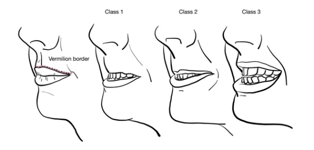
Report: I note normal mandibular protrusion, and a normal Class 1 Upper Lip Bite Test.
Why Check: Part of the LEMON mnemonic used to assess for a difficult airway. The Upper Lip Bite test is strongly validated independently to assess whether the patient will be difficult to intubate. Class 1 (normal) is where the lower incisors extend beyond vermillion border; Class 2 is where lower incisors bite upper lip before vermillion border; and Class 3 is where lower incisors cannot bite upper lip at all. Class 2 and above on upper lip bite test greatly increases the risk of difficult intubation (LR+ 12, LR- 0.42). The Mandibular protrusion test is part of Wilson Score, and independently moderately increases likelihood of a difficult intubation (LR+ 5.5)
3-3-2 Rule (LEMON: Evaluate)
Say: Can you please look straight ahead and open the mouth as wide as you can?
Do: Ask patient to try to place 3 of their fingers in between upper and lower incisors.
Say: Thank you. Now, can you please close the mouth?
Do: Ask patient to try to place 3 fingers along mandible (hyomental distance – hyoid bone to chin). Then, ask patient to place 2 fingers in the superior laryngeal notch (hyothyroid distance – thyroid notch to hyoid bone).
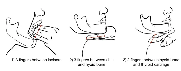
Report: I note a normal 3-3-2 rule.
Why Check: Part of the LEMON mnemonic used to assess for a difficult airway. This helps to assess the alignment of pharyngeal, laryngeal and oral axes to assess whether the patient will be difficult to intubate. A hyomental distance of <5.5 cm moderately increases the likelihood of difficult intubation (LR+ 6.4, LR- 0.84). If unable to use patient’s own fingers, using a gloved hand you can try to use your own fingers here.
Thyromental Distance (TMD) (LEMON: Evaluate)
Say: Can you please sit up straight, and look up as far as you can comfortably?
Do: Measure the distance between the thyroid notch and the mentum (chin).
Report: The patient has a thyromental distance of ≥7cm.
Why Check: Part of the LEMON mnemonic used to assess for a difficult airway. The TMD spans from the mentum to the thyroid notch. This distance encompasses the hyomental distance and the hyothyroid distance (from the 3-3-2 rule). The 3-3-2 rule assesses a neutral neck, while the TMD assesses a neck in full extension. A short TMD means the laryngeal axis is in a more acute angle with the pharyngeal axis. A height:TMD ratio >17 moderately increases the likelihood of difficult intubation (LR+ 5.2, LR- 0.36), and short distance (<7cm) mildly increases the likelihood of a difficult intubation (LR+ 3.3, LR- 0.63)
Mallampati Score (LEMON: Mallampati)
(Also seen in Ch 7 – Head and Neck Exam – MF1)
Say: I will inspect the mouth to look at airway structures. Can you briefly open the moth and stick the tongue out?
Do: Inspect the patient’s oropharynx structures for the soft palate, uvula, fauces, and pillars.
Scoring: see image. Mallampati 1 = soft palate, uvula, pillars completely visible. Mallampati 2 = soft palate and part of uvula visible. Mallampati 3 = only soft palate and base of uvula visible. Mallampati 4 = soft palate not visible.
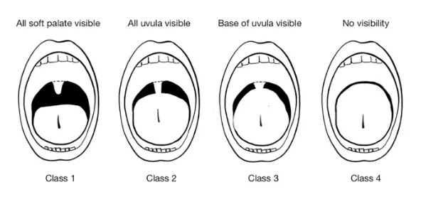
Report: I can visualize all of the soft palate, uvula and pillars. The Mallampati Score is 1.
Why Check: Part of the LEMON mnemonic used to assess for a difficult airway. Mallampati Class I or II classifies a patient’s airway as easier, and Mallampati Class III or IV classifies an airway as difficult, where a Mallampati Class III or IV airway mildly increases the likelihood of a difficult intubation (LR+4.1, LR-0.52). A Mallampati Score of 3 or 4 is also significant in assessing patients for Obstructive Sleep Apnea.
Cervical Spine Range of Motion (ROM) (LEMON: Neck)
(Also seen in Ch 29 -Back Exam – MF4)
AFMC level Pre-clerkship 3 · Clerkship 4
Say: I will now assess for active neck mobility. Could you sit up straight, and look straight ahead? Please look in each direction until you feel discomfort or cannot look further.
Do – Active ROM: Assess the patient from their right side (i.e. looking at patient laterally in profile as they form a plane flexing and extending their C-spine). Ask patient to look up (neck extension) as far as they can. If available, place a marker vertically against the patient’s forehead. Then ask the patient to look down (neck flexion) as far as they can, trying to have chin touch their chest. Measure the angle formed between the marker when neck fully extended versus fully flexed.
Report: The patient’s cervical spine is normal on active range of motion in flexion and extension.
Why Check: Part of the LEMON mnemonic used to assess for a difficult airway, and also a part of the Wilson Score. The cut-off point in this test is 90 degrees. Normal C-Spine ROM includes flexion to chin touching chest (80-90°) and extension to chin should be past level of ears (70°). If concerned that active ROM is limited by pain/discomfort, you can also assess passive ROM. DO NOT assess if concern for unstable C-Spine (e.g. Canadian C-Spine Rule, NEXUS Criteria, patient in hard collar).
Wilson Score
Do: Using results of parameters obtained above, calculate the patient’s Wilson Score, as follows:
| Parameter | + 0 Points | + 1 Point | + 2 Points |
|---|---|---|---|
| Weight (kg) | < 90 | 90-110 | > 110 |
| Cervical Spine Mobility | > 90° | 90° | < 90° |
| Impaired Jaw Mobility | Inter-incisor Gap ≥ 5cm or lower teeth can protrude past upper teeth | Inter-incisor Gap < 5cm and lower can meet upper teeth | Inter-incisor Gap < 5cm and lower can’t meet upper teeth |
| Retrognathia | Normal | Moderate | Severe |
| Prominent Incisors | Normal | Moderate | Severe |
Report: I note the patient’s Wilson Score is 1 point, for Weight 100kg.
Why Check: A Wilson Score ≥ 2 moderately increases the likelihood of a difficult intubation (LR+ 9.1, LR- 0.60). Individual components of airway assessment are not as sensitive or specific for difficult intubation as a combined assessment here.
Cormack-Lehane Grade
Do: During direct laryngoscopy only, assess the view. Characterize as follows:
- Grade 1 - Full view of glottis.
- Grade 2a – Partial view of glottis.
- Grade 2b – Only posterior extremity of glottis or arytenoid cartilages seen.
- Grade 3 – Only epiglottis seen, no glottis.
- Grade 4 – no glottis/epiglottis seen.
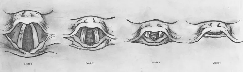
Cormack-Lehane Grade18
Report: On direct laryngoscopy, I am able to obtain a Grade 1 view and can successfully intubate the trachea.
Why Check: Grades the adequacy of the view of the larynx during direct laryngoscopy. Helps ensure the view is adequate (Grade 1-2) prior to intubation to ensure successful tracheal intubation. Grade 3-4 is the definition of “difficult” airway.
Focused Inspection
General Appearance
AFMC level Pre-clerkship 4 · Clerkship 4
Say: I will begin with general inspection.
Do: Attend to the patient’s general appearance throughout the interview.
Report: I observe the patient as a frail, elderly gentleman with cachexia, ambulating with a walker, who appears older than his stated age, who is alert and oriented, and is calm and cooperative.
Why Check: The general appearance of a patient can shed light on their general functional status and overall clinical picture and guide focused exams of particular body systems in the pre-op assessment. Their mental status examination (See Ch 32 – Approach to Psychiatric Consultation – MF4) can also influence anesthetic approach (e.g. pre-medication for anxiety; general vs. regional anesthesia approach). Medicine is an art of attentive observation, and your sense of the patient’s general appearance can be honed during the interview.
Central Cyanosis
AFMC level Pre-clerkship 4 · Clerkship 4
Say: I will now inspect under the tongue for signs of low oxygen levels. Can you briefly open the mouth, and lift the tongue up?
Do: Inspect under the tongue for central cyanosis
Report: I note no signs of central cyanosis; I note the tongue appears proper size, free in motion, slightly red in colour and with thin white coating layer.
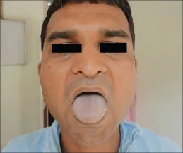
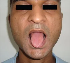
Central Cyanosis19 and Cyanosis Resolved19
Why Check: Central cyanosis indicates poor oxygenation in the lungs (specifically PaO2 <50mmHg given an Hb of 140-160g/L; PaO2 45mmHg if Hb 120 g/L).
Work of Breathing
AFMC level Pre-clerkship 4 · Clerkship 4
Say: If you can breathe normally, I will examine the face and torso for signs of work of breathing.
Do:
- Look for signs of increased work of breathing: tripod position, pursed lips, nasal flaring, accessory muscle use (sternocleidomastoid, scalene, trapezius, internal intercostal, or abdominal muscles), paradoxical breathing (Hoover’s sign), tracheal tug (abnormal downward movement of trachea during inspiration), suprasternal indrawing, intercostal indrawing, supraclavicular retraction, grunting (pediatrics).
- Observe if patient can finish complete sentences (not interrupted by work of breathing).
Report: There are no signs of increased work of breathing (comment on all the above out loud as you observe on inspection). You are speaking in complete sentences which is reassuring as well.
Why Check: Important in assessing dyspneic patients and assessing if a patient is in respiratory distress. The inability to speak in complete sentences demonstrates a higher degree of respiratory distress.
Clubbing (Schamroth Sign)
AFMC level Pre-clerkship 4 · Clerkship 4
Say: I will now inspect the fingers for signs of low oxygen levels. Can you hold the index finger nails together like this? (See picture, model for patient)
Do: Model with your own hands. Look for a visible gap between the two nail beds.
- If arthritic changes, check for a concave bump between base of nail bed and finger
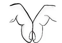
Report: Schamroth’s sign is negative, with no evidence of clubbing.
Why Check: Associated with chronic hypoxia, cyanotic heart disease, lung cancer, interstitial lung disease
Peripheral Cyanosis
AFMC level Pre-clerkship 4 · Clerkship 4
Say: I will now inspect the hands for signs of changes in circulation. Can you show the hands with palms up? And now with palms down?
Do: Hold hands lightly and inspect for peripheral cyanosis
Report: I note no signs of peripheral cyanosis.
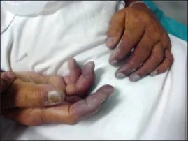
Peripheral Cyanosis20
Why Check: Peripheral cyanosis indicates inadequate circulation
Focused Palpation
PMI (Point of Maximal Impulse)
- Loc.– [3, 4]
- Amp. – [1, 1]
- Dur. – [1, 2]
- Size – [2, 4]
Say: I will palpate the chest for where the heart beats strongest. Can you lean forward?
Do: Using 2-3 fingers, locate the 5th intercostal space at left mid-clavicular line (MCL) – the expected site of PMI. Assess Location, Amplitude, Duration and Size (LADS).
Report: PMI located at 5th intercostal at left MCL, is brisk and tapping, lasts 2/3rd systole, and size of quarter.
Why Check: Apex beat palpation can provide evidence of hypertrophy – PMI not always apical impulse
- Location of Apex: Displaced location can be evidence of decreased ejection fraction, left ventricular dilatation, increased left atrial pressure, or enlarged Cardiothoracic Ratio on Chest X-Ray.
- Amplitude of Apex: If patient has known mitral stenosis, increased apex amplitude suggests comorbid aortic stenosis or mitral regurgitation
- Duration of Apex: Sustained apex with an aortic murmur argues for aortic stenosis; Lack of sustained apex with Aortic Regurgitation argues against moderate-severe severity of Aortic regurgitation
- Size of Apex: Increased size evidence of increased LV End-Diastolic Volume
Peripheral Venous Access Assessment
AFMC level Pre-clerkship 2 · Clerkship 4
Say: I am now going to check the left arm for planning which veins we can access during the surgery.
Do: Assess Peripheral Venous Access sites – distally at dorsal venous arch (have patient grasp your hand while arm pronated, pull with gentle traction on MCPs by thumb to expose veins); transition patient arm to supination; assess radial and ulnar veins; in antecubital fossa, cephalic, median antecubital, and basilic veins; and proximally, the axillary vein. You can gently tap on veins to cause vasodilation; and can also apply a temporary tourniquet to occlude veins proximal to tourniquet to assess distal venous sites). Look for previous axillary dissection/edema, previous AV fistula for Hemodialysis that could impact site use.
Report: Based on the planned surgery, I recommend we start a 20 Gauge IV at the Left cephalic vein.
Why Check: Typically, an anesthesiologist is positioned to patient’s left side with left arm generally used. IV site choice depends on venous anatomy; intended rate requirements (vein size limits max tubing radius of IV which affects max flow rate to fourth power – Poiseuille’s Law); intended duration of use; and patient preference.
*Note: Central Venous Catheters (internal jugular, subclavian, femoral veins) may be placed – indications include inadequate peripheral IV access, peripherally incompatible meds (e.g. infusions of vasopressors, chemotherapy, Total Parenteral Nutrition = TPN), hemodynamic monitoring, and extracorporeal therapy (e.g. hemodialysis). Inserting peripheral IVs and central lines is an important skill you encounter in Clerkship.
Focused Auscultation
Listening to Breathing
(Also seen in Ch 5 – Resp Exam)
- General- [3, 4]
- Stridor – [4, 4]
- Pleural Rub- [1, 4]
- Stridor (Peds) – [1, 3]
- Wheeze (Infant) – [1, 2]
Say: I will now listen to the chest and back – breathe in and out each time you feel the stethoscope. Please pause if you become light-headed.
Setup: Patient crosses arms to displace scapulae.
Do: Hold stethoscope still when listening (don’t want to only hear rustling of moving it around).
Report: I note air entry bilaterally with vesicular breath sounds and no adventitious sounds.
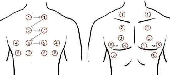
What is Normal? Normal auscultation depends on listening location
- (1) Trachea: Bronchial – harsh, high-pitched, heard through full inspiration and expiration
- (2) Sternum: Bronchovesicular – less harsh, heard through full inspiration, short and softer on expiration
- (3) Everywhere else: Vesicular - gentle, low-pitched, quiet on inspiration and nearly silent on expiration
Why Check: Respiratory auscultation is important to assess for respiratory comorbidities in the context of anesthetic planning. In particular, listen for Adventitious (Abnormal) Sounds, including:
| Sound | Respiratory Phase | Description | Significance |
|---|---|---|---|
| Fine Crackles | Inspiration | Discontinuous, soft, high-pitched, more per breath, popping sound. | Distal lower airway process (e.g. pulmonary fibrosis, congestive heart failure) |
| Coarse Crackles | Inspiration alone; or Inspiration + early expiration | Discontinuous, loud, low-pitched, fewer per breath, bubbling sound. | Proximal lower airway process (e.g. pneumonia, pulmonary edema, adult respiratory distress syndrome) |
| Stridor | Inspiration | Loud, musical sound, constant pitch, louder over neck. | Upper airway obstruction (e.g. croup, foreign body aspiration, epiglottitis, bacterial tracheitis, retropharyngeal abscess, peritonsillar abscess) |
| Pleural Rub | Expiration alone; or Expiration + Inspiration | Loud, grating or rubbing sound; can be crackling. | Pleural inflammation (e.g. pleural effusion, pulmonary embolism, pleuritis) |
| Wheeze | Expiration alone; or Expiration + Inspiration | Continuous, high-pitched, musical sound. | ↓ Distal lower airway diameter (e.g. asthma, chronic obstructive pulmonary disease) |
| Rhonchus | Expiration alone; or Expiration + Inspiration | Continuous, low-pitched, coarse sound; may clear after cough. | ↓ Proximal lower airway diameter by mucous secretions, lesions or foreign bodies (e.g. pneumonia, cystic fibrosis) |
-
Bronchial breath sounds over lung (normal over trachea) points to consolidation
-
Diminished breath sounds greatly increases likelihood for suspected pleural fluid (LR+ 18.6)
-
Early Crackles increases likelihood of suspected obstructive disease (LR+ 14.6), and particularly for severe obstruction in patients with chronic airflow limitation (LR+ 20.8; LR- 0.1)
-
Stridor: Sensitivity of 80.4% & specificity of 59.5% for Foreign Body Aspiration21. May be the earliest sign worrisome for airway compromise.
-
Note - Pediatrics: Wheezing in Infants: Respiratory exam of wheezing in infants shows good inter-rater reliability, but respiratory status overall is most reliable22.
-
Issue: Adventitious sounds on auscultation is “the most ambiguous and confusing nomenclature in all of physical diagnosis” where clinicians use diverse terms to describe similar sounds23.
-
Modification for Patients with Limited Mobility or Supine Patients: For auscultation of the lungs, auscultate anteriorly, then roll onto a lateral decubitus position to auscultate lung bases posteriorly.
-
Modification for Patients with Obesity: Auscultate directly over exposed skin, to ensure breath sounds can be appreciated.
Cardiac Precordial Auscultation
(Also seen in Ch 6 – Cardiac Exam)
- S3, S4- [1, 3]
- Murmur [1, 3]
- Abnormal S1, S2 [1, 1]
- Position [1, 4]
Say: I will auscultate the chest for heart sounds. Please breathe normally as I listen.
Do: Listen carefully with the diaphragm of the stethoscope at each valve region (Aortic, Pulmonic, Tricuspid, and Mitral). Listen for abnormal heart sounds or murmurs.
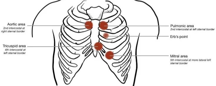
Report: I note a normal S1 and S2. There are no extra heart sounds or murmurs present.
What Is Normal? S1 and S2, regular rate and rhythm, with no extra heart sounds and no murmurs
- S1 = loudest at Tricuspid & Mitral valves (re atrioventricular valves closing at start of systole)
- S2 = loudest at Aortic & Pulmonic valves (re semilunar valves closing at end of systole). Physiologic splitting of S2 is normal on inspiration due to ↑ negative intrathoracic pressure – best heard in a sitting position.
Why Check: Cardiac auscultation important to assess for cardiac comorbidities as part of anesthetic planning
Abnormal Findings – Abnormal S1 and S2 and Associated Conditions:
| Characteristic | S1 | S2 |
|---|---|---|
| Loud | Mitral Valve Prolapse (MVP); Mitral Stenosis (MS); Left Atrial Myxoma; Tachycardia | Systemic hypertension (loud A2) or Pulmonary hypertension (loud P2) |
| Faint | Mitral Regurgitation; Left Bundle Branch Block (LBBB); Long PR interval (1st degree AV block) Acute Aortic Regurgitation (AR) | Aortic Stenosis (LR+ 7.5, LR- 0.5) |
| Variable | AV Dissociation (LR+ 24.4, LR- 0.4); Atrial Fibrillation | N/A |
| Wide Split | Right Bundle Branch Block (RBBB); Right Atrial Myxoma | RBBB; Pulmonic Stenosis; Mitral Regurgitation; Ventricular Septal Defect (VSD); Right Ventricular Failure |
| Paradoxical Split | N/A | LBBB; Severe Aortic Stenosis; Wolff-Parkinson-White (WPW); LV Dysfunction |
| Fixed Split | N/A | Atrial Septal Defect (ASD) |
Abnormal Findings – Extra heart sounds: Best heard in left lateral decubitus position.
- S3: Heard just after S2, with rapid and early diastolic filling of ventricles, “Sloshing in” (e.g. volume overload). In the appropriate clinical context (e.g. dyspneic patients in Emergency Department), hearing an S3 increases the likelihood of Congestive Heart Failure (LR+11, LR- 0.88).
- S4: Heard just before S1, with diastolic rapid late filling of ventricles, “A stiff wall” (e.g. pressure overload, Left Ventricular Hypertrophy).
- S3 + S4: Summation gallop
Abnormal Findings – Murmurs (represent Turbulent blood flow). Listen for Location (i.e. identify valve); Radiation (e.g. to carotids, left sternal border, etc.); Timing (e.g. Systolic vs. Diastolic); Configuration (e.g. Crescendo, Decrescendo, Crescendo-decrescendo, Plateau); Quality (e.g. high vs. low pitch; harsh, musical, blowing, rumbling, etc.); Change with maneuvers (e.g. with inspiration, Valsalva, squat); and Volume/Grade
- Systolic Murmur: Grades 1 (very faint); Grade 2 (soft, readily detectable); Grade 3 (easily detectable without thrill); Grade 4 (easily detectable with thrill); Grade 5 (very loud murmur audible with stethoscope placed lightly on chest); Grade 6 (extremely loud, audible with stethoscope off chest)
- Diastolic Grade: Grade 1 (barely audible); Grade 2 (audible, soft); Grade 3 (easily audible); Grade 4 (loud)
Mitral Stenosis (MS)
- Pathophysiology: Mitral valve should be open in diastole – mitral stenosis causes incomplete opening of mitral valve in diastole
- Murmur: Diastolic murmur with Opening Snap best heard at cardiac apex in left lateral decubitus position. Worsened by ↑ venous return (squat/leg raise)
Aortic Regurgitation / Insufficiency (AR/AI)
- Pathophysiology: Aortic valve should be closed in diastole – aortic regurgitation causes incomplete closing of aortic valve in diastole
- Murmur: Diastolic murmur heard best at right base in sitting position. Worsened by ↑ venous return (squat/leg raise)
Aortic Stenosis (AS)
- Pathophysiology: Aortic valve should be open in systole – aortic stenosis causes incomplete opening of aortic valve in systole
- Murmur: Systolic murmur with crescendo-decrescendo pattern heard best at right base. Radiates to R clavicle and/or R carotid. Worsened by ↑ venous return (squat/leg raise)
Hypertrophic Cardiomyopathy (HCM)
- Pathophysiology: LV Outflow Tract Obstruction due to sarcomere gene mutations.
- Murmur: Systolic murmur with crescendo-decrescendo pattern heard best at cardiac apex and left lower sternal border. May radiate to axilla. Improved by ↑ venous return (squat/leg raise)
Mitral Regurgitation (MR)
- Pathophysiology: Mitral valve should be closed in systole – mitral regurgitation causes incomplete closing of mitral valve in systole.
- Murmur: Systolic murmur with holosystolic pattern heard best at cardiac apex. Radiates to axilla. Worsened by ↑ venous return (squat/leg raise)
Mitral Valve Prolapse (MVP)
- Pathophysiology: Mitral valve should be closed in systole, where thickened mitral valve leaflets in MVP cause imperfect closure of mitral valve in systole.
- Murmur: Systolic murmur similar to Mitral regurgitation, but improved by ↑ venous return (squat/leg raise)
Maneuvers to Accentuate/Differentiate Murmurs:
| Maneuver | RV Preload | LV Preload | LV Afterload | Effect on Murmurs |
|---|---|---|---|---|
| Inspiration | ↑ | ↓ | ∅ | ↑ R-sided murmurs, ↓ L-sided murmurs (except HCM) |
| Valsalva (Strain Phase)/ Abrupt Stand | ↓ | ↓ | ↓ | ↑ MVP/HCM, ↓ MS/MR/AS/AR/VSD |
| Squatting/Leg raise | ↑ | ↑ | ∅/↑ | ↓ MVP/HCM, ↑ MS/MR/AS/AR/VSD |
| Hand Grip (for 20-30s) | ∅ | ∅ | ↑ | ↓ MVP/HCM; ↑ MR/AR/VSD; ↓ MS/AS |
Note on Pediatric Cardiac Auscultation: Not all pediatric murmurs are pathologic; pathology more likely if murmur is systolic grade 3+; holosystolic; associated systolic click; diastolic; worse on standing; or harsh.
Modification for Patients with Obesity: Ask the patient to lean forward if sitting, or to place their above their head if supine. Leaning forward brings the heart closer to the chest wall & placing arms above the head when supine redistributes the soft tissue of the chest wall, making heart sounds easier to appreciate.
Part 4: Select additional exams that may be necessary depending on comorbidities identified
In previous MFs, students have practiced focused physical exams which may be required as part of the Pre-Operative Anesthesia assessment, depending on comorbidities identified and anesthesia planning considerations. Examples include:
Respiratory Exam (Ch 5 – MF1)
- Inspection:
- Barrel Chest (associated with hyperinflation) and Flail Chest (sign of rib fractures at two points)
- Pectus Excavatum and Carinatum (can impair respiratory and cardiac function if severe)
Cardiac Exam (Ch 6 – MF1), Peripheral Vascular Exam (Ch 8 – MF1) and Volume Status Exam (Ch 9 – MF2)
- Inspection: Jugular Venous Pressure (re. intravascular volume status)
- Palpation:
- Edema (pitting edema may be due to increased hydrostatic pressure e.g. congestive heart failure, or due to decreased oncotic pressure e.g. in nephrotic syndrome, hypoalbuminemia; non-pitting edema may be exudate-filled, or due to lymphedema).
- Peripheral pulses (re. signs of arterial insufficiency)
- Central pulses (re. signs of obstructive arterial pathologies)
Abdominal Exam (Ch 13 – MF3)
- Assessment for Liver Cirrhosis: Liver cirrhosis may impact drug metabolism, fluid status and coagulation status intraoperatively, as an important comorbidity to assess if present – e.g. assess for ascites, hepatosplenomegaly, and stigmata of liver disease.
- Assessment for Peritonitis: Involuntary Guarding/Rigidity and Rebound Tenderness on palpation.
Back Exam (Ch 29 – MF4)
- Key Idea: Palpating for bony landmarks is especially important when planning neuraxial anesthesia.
- Palpation: Assess postural alignment (Normal: cervical lordosis, thoracic kyphosis, lumbar lordosis, sacrococcygeal kyphosis) including for kyphoscoliosis. Palpate from occiput inferiorly for spinous processes (including C7 – prominens at base of neck; T3 – spine of scapula; T7 – angle of scapula; L3/L4 – iliac crests) and anterior superior iliac spine (ASIS).
Screening Neurological Exam (Ch 31 – MF4)
- Key Idea: Important to assess in patients with history of pre-existing motor or sensory deficits. For regional nerve block anesthetic approaches, it is important to assess for deficits at baseline to be able to differentiate from post-operative deficits should they occur.
- Motor Exam: Assess for gross motor deficits, symmetry, tone, posturing and abnormal movements.
- Sensory Exam: Assess for posterior column and spinothalamic tracts.
Part 5: Special Population Considerations – Pediatrics and Obstetrics
Pediatric Population
Pediatric patients are a unique patient population with additional considerations during the preoperative assessment. In addition to the above history and physical exam maneuvers, the following should be considered on history:
Focused SSHADESS: Adolescent Interview
- General Approach: Begin with a confidentiality statement and speak to the adolescent privately without caregivers in the room. An example confidentiality statement is as follows:
- As with all of our visits, the information we discuss will be kept confidential. I might take notes and put those in your record here, but everything will be stored securely. There are some instances where I may be required by law to break confidentiality, for example if I believe there is immediate risk of harm to yourself or others. Does this sound okay with you?
- Focus specifically on:
- Drugs/substance use: Do any of your friends smoke (cigarettes, vaping, marijuana, etc.) use alcohol or other drugs? Have you ever tried these things? How much do you use and how often? What situations do you use those substances in? Do your family members use drugs, including alcohol and tobacco? Do you use any performance enhancing drugs?
- Sexuality: What is your name? What are your pronouns? Have you ever been in a relationship? Who are you attracted to? What kind of sex have you engaged in? How many partners have you had? Do you use contraception and/or protection? Have you ever been pregnant? Do you have any symptoms or concerns about STIs?
- Other Elements of SSHADESS Adolescent Interview: Strengths; School; Home; Activities; Eating/Emotions; and Safety.
Why Check: Recall that capacity to consent for treatment is not age-dependent in Ontario, where the physician must make a determination of capacity for a minor as they would for an adult. If the minor is capable to provide consent to a treatment, the physician must obtain consent from the minor directly, even if the minor is accompanied by a parent or guardian24. As per the Canadian Pediatric Society, “Children’s and adolescents’ participation in medical decision-making should be sought in proportion to their developmental capacity to understand the nature and consequences of their medical problem as well as to reasonably foreseeable risks and benefits of the treatment performed” where “health care providers, patients and families should work together to reach medical decisions based on the patient’s best interests or outcomes” within a family-centred, shared decision-making model25.
As patients become adolescents, it is important to normalize offering part of the assessment occur with the adolescent alone, including the SSHADESS interview (e.g. offer to perform part of the history with the adolescent alone, then invite the parent or guardian back into the room for the physical examination or creating a management plan). This may also give space for the adolescent to express their understanding and appreciation of the proposed surgical treatment, its risks, benefits and alternatives and likely consequences of not having the treatment.
Focused Birth History
In particular, focus on:
- Antenatal history/family medical history: History of malignant hyperthermia, pseudocholinesterase deficiency?
- Prenatal screening/newborn screening tests: Any positive tests?
- Prematurity: Number of weeks gestation – Pre-term <37 wk; Early term: 37wk – 38wk + 6d; Full Term: 39wk – 40wk + 6d; Late Term: 41wk – 41wk + 6d; Post-Term ≥42 wk 0d
Why Check: Prematurity can affect anesthetic management and lead to additional complications such as bradycardia and apnea up to 24 hours after surgery. Pediatrics patients account for 52.1% of all cases of malignant hyperthermia. Identifying at-risk children is key in preventing its occurrence and will affect the anesthetic plan. Identifying congenital or other disorders will likewise affect anesthetic agents used and the anesthetic plan.26
Addressing child/parental anxiety
- Provide education to caregivers/patient regarding likelihood of common risks; adverse event risk in healthy child undergoing uncomplicated surgery very low (1/200,000).25
- Discuss induction day plan.
Why Check: The perioperative period is highly stressful for caregivers and pediatric patients. RCTs show that caregiver presence in the OR during induction is generally not beneficial; anxious caregivers may increase anxiety levels in the child. Other options to reduce perioperative anxiety include caregiver/patient education, involvement of child life specialists in the OR, topical freezing for IV insertion, oral anxiolytics like midazolam, and tours of the OR and PACU prior to surgery. Identifying and addressing caregiver anxiety is important, as it is a predictor for increased anxiety in pediatric patients. High levels of anxiety in the perioperative period are linked to poorer outcomes such as increased postoperative pain, delirium, and behaviour changes.262728
Obstetrics Population
Obstetrical patients have unique considerations. There are numerous physiologic changes that occur during pregnancy, including but not limited to:
- Cardiovascular: Increased blood volume (due to hormonal effects), increased cardiac output (due to ↑ metabolic demand)
- Respiratory: Mucosal edema (increased risk of difficulty with tracheal intubation and bleeding with airway instrumentation), increased minute ventilation and tidal volume (due to increased O2 consumption); decreased functional reserve capacity (due to rising uterus displacing diaphragm); increased PaO2 by 10mmHg (due to hyperventilation)
- Gastrointestinal: Increased gastric fluid volume (due to rising uterus displacing pylorus and delaying gastric emptying); increased gastric fluid acidity (due to gastrin secretions by placenta)
In addition, neuraxial anesthetic techniques are increasingly common as part of intrapartum care. The following should be considered in obstetrical patient populations, in addition to the above history questions and exam maneuvers.
Focused obstetrical history
- Maternal Health History:
- Pre-existing medical conditions
- Maternal medications
- Maternal substance use (smoking, EtOH, marijuana, IVDU)
- Maternal blood type; Hx of previous child w/ ABO or Rh incompatibility
- Obstetrical History and Current Pregnancy:
- GTPAL (G = gravidity; T = term; P = preterm; A = abortions; L = living)
- Past Pregnancies: Year, sex, weight, gestational age, preterm, preterm rupture of membranes, need for induction, length of labour, method of delivery (if C-section, ask why), complications
- Past history of complications: Gestational diabetes mellitus (GDM), hypertensive disorders of pregnancy (e.g. gestational hypertension, pre-eclampsia, HELLP syndrome = Hemolysis, Elevated Liver Enzymes, Low Platelets), thrombocytopenia.
- Key Question: Any complications during this pregnancy?
Oral intake: Last time patient ate/drank
Why Check: Obstetrical complications such as HELLP syndrome and thrombocytopenia can contribute to anesthetic complications. In patients who may be high risk for operative deliveries, additional restrictions on PO intake may be necessary.29
Focused physical exam maneuvers
- Baseline BP check
- Fetal heart rate (FHR) monitoring – note that for Epidural analgesia, Intermittent Auscultation (IA) may continue to be used in low-risk term pregnancies with spontaneous labour; for Combined spinal-epidural analgesia, Electronic Fetal Monitoring (EFM) is recommended due to increased risk of atypical or abnormal FHR patterns29.
- In the case of neuraxial anesthesia, perform a focused back exam: ROM (should be able to sit in ‘cat arching back’ position), inspect for infection around the site of needle insertion, palpate for scoliosis or other deformities, and inquire about prior back surgeries or hardware in place.
Why Check: Blood pressure (BP) and fetal heart rate (FHR) can be influenced by anesthetic agents. FHR should be monitored before and after regional anesthesia. Neuraxial anesthesia can drop maternal BP, leading to changes in FHR and should also be monitored, along with O2 sats.3031 Note that epidural analgesia may be expected to add up to 1 hour in the 2nd stage passive phase of labour (cervix is fully dilated, patient is not actively pushing).
Part 6: Relevant Likelihood Ratios – from Rational Clinical Examination JAMA series
Clinical Question 1: Will this patient be difficult to intubate?32
Definition of “Difficult”: Cormack-Lehane Grade 3-4 on Direct laryngoscopy
- Grade 1 - Full view of glottis
- Grade 2a – Partial view of glottis
- Grade 2b – Only posterior extremity of glottis or arytenoid cartilages seen
- Grade 3 – Only epiglottis seen, no glottis
- Grade 4 – neither glottis nor epiglottis seen
| Risk Factors | LR+ (95% CI) | LR- (95% CI) |
|---|---|---|
| History of difficult intubation | 16-19 (about +45%) | 0.72-0.82 (about −15%) |
| Difficult Bag-Mask Ventilation prior to intubation | 3.5 (2.6-4.7) (about +15%) | 0.67 (0.55-0.80) (about −15%) |
| History of Snoring | 3.4 (1.6-7.3) (about +15%) | 0.65 (0.58-0.72) (about −15%) |
| Overweight/Obesity (BMI >27-35) | 2.2 (1.6-3.1) (about +15%) | 0.70 (0.46-1.1) (no change: 95% CI includes 1) |
| Male (vs. Female) | 1.2 (1.0-1.3) (no change: 95% CI includes 1) | 0.87 (0.76-0.99) (about −15%) |
| Physical Examination | Sensitivity | Specificity | LR+ (95% CI) | LR- (95% CI) |
|---|---|---|---|---|
| Upper Lip Bite Test – Class 3 | 0.60 (0.42-0.76) | 0.96 (0.93-0.97) | 14 (8.9-22) (about +45%) | 0.42 (0.27-0.65) (about −30%) |
| Upper Lip Bite Test – Class 2 or 3 | 0.60 (0.46-0.73) | 0.95 (0.91-0.97) | 12 (6.9-20) (about +45%) | 0.42 (0.30-0.59) (about −30%) |
| Wilson Score ≥ 2 | 0.43 (0.26-0.62) | 0.95 (0.90-0.98) | 9.1 (5.1-16) (about +30%) | 0.60 (0.44-0.82) (about −15%) |
| Hyomental Distance (<3 - <5.5cm) | 0.20 (0.11-0.34) | 0.97 (0.94-0.98) | 6.4 (4.1-10) (about +30%) | 0.84 (0.73-0.96) (about −15%) |
| Retrognathia (Mandible <9cm, or subjectively short) | 0.19 (0.07-0.42) | 0.98 (0.90-1.0) | 6.0 (3.1-11) (about +30%) | 0.85 (0.76-0.94) (about −15%) |
| Impaired mandibular protrusion (unable to move lower teeth past upper teeth) | 0.25 (0.06-0.63) | 0.95 (0.86-0.99) | 5.5 (2.1-15) (about +30%) | 0.78 (0.54-1.1) (no change: 95% CI includes 1) |
| Height:Thyromental distance ratio (≥17 - ≥25) | 0.69 (0.57-0.78) | 0.87 (0.67-0.95) | 5.2 (1.9-14) (about +30%) | 0.36 (0.25-0.52) (about −30%) |
| Thyromental distance (<4 - <7 cm) | 0.45 (0.36-0.55) | 0.86 (0.80-0.91) | 3.3 (2.4-4.4) (about +15%) | 0.63 (0.55-0.73) (about −15%) |
| Impaired neck mobility (<30-90° flexion/extension, or subjective assessment) | 0.28 (0.13-0.51) | 0.93 (0.85-0.97) | 4.2 (1.9-9.5) (about +15%) | 0.77 (0.60-0.99) (about −15%) |
| Sternomental distance (<12-15cm) | 0.41 (0.27-0.57) | 0.90 (0.83-0.94) | 4.1 (2.7-6.1) (about +15%) | 0.65 (0.52-0.82) (about −15%) |
| Modified Mallampati Score ≥ 3 | 0.55 (0.48-0.62) | 0.87 (0.81-0.91) | 4.1 (3.0-5.6) (about +15%) | 0.52 (0.45-0.60) (about −15%) |
| Impaired mouth opening (Inter-incisor gap <2-5cm) | 0.36 (0.20-0.56) | 0.90 (0.80-0.95) | 3.6 (2.1-6.1) (about +15%) | 0.71 (0.55-0.92) (about −15%) |
| Palm print (varies on scoring system for each article) | 0.77 (0.22-0.98) | 0.84 (0.55-0.96) | 3.0 (1.9-4.7) (about +15%) | 0.28 (0.08-0.97) (about −30%) |
| 3-3-2 Rule (Score ≥2) | 0.66 (0.53-0.77) | 0.69 (0.65-0.72) | 2.1 (1.7-2.6) (about +15%) | 0.50 (0.36-0.70) (about −15%) |
Clinical Question 2: Will this patient be difficult to Bag-Mask Ventilate (BMV)?33
Definition of “Difficult”:
- Inability to maintain O2sat >92% with 100% O2 and Positive Pressure Mask Ventilation (BMV)
- Important gas flow leak by face mask
- Necessity to ↑ Flow to >15L/min and use Oxygen Flush Valve >2 times
- No perceptible chest rise
- Necessity to use 2-handed BMV technique
- Change of operator required.
| Risk Factors | OR+ (95% CI) | P-Value |
|---|---|---|
| B – Beard | 3.18 (1.39-7.27) | 0.006 |
| O – Overweight/Obesity (BMI >26 kg/m²) | 2.75 (1.64-4.62) | <0.001 |
| N – No Teeth | 2.28 (1.26-4.10) | 0.006 |
| E – Elderly (Age > 55yo) | 2.26 (1.34-3.81) | 0.002 |
| S – Snoring History | 1.84 (1.09-3.10) | 0.02 |
| Number of BONES Risk Factors | Sensitivity | Specificity | PPV | NPV |
|---|---|---|---|---|
| 1 | 0.92 | 0.38 | 0.07 | 0.99 |
| 2 | 0.72 | 0.73 | 0.12 | 0.98 |
| 3 | 0.35 | 0.91 | 0.17 | 0.96 |
| 4 | 0.07 | 0.99 | 0.24 | 0.95 |
| 5 | 0.00 | 1.00 | 0.00 | 0.95 |
References
-
Mocon A, et al. Drop The Pre-Op: A toolkit for reducing unnecessary visits and investigations in pre-operative clinics. Version 1.1. Choosing Wisely Canada; 2019. ↩ ↩2 ↩3 ↩4 ↩5
-
Recommendations. Choosing Wisely Canada. Accessed August 1, 2022. https://choosingwiselycanada.org/recommendations/#list ↩
-
American Society of Anesthesiology. Practice guidelines for preoperative fasting and the use of pharmacologic agents to reduce the risk of pulmonary aspiration. Anesthesiology. 2017;3(126):376-393. ↩
-
Duceppe E, et al. Canadian Cardiovascular Society guidelines on perioperative cardiac risk assessment and management for patients who undergo noncardiac surgery. Can J Cardiol. 2017;33(1):17-32. ↩ ↩2 ↩3 ↩4
-
Global Initiative for Chronic Obstructive Lung Disease (GOLD). Global Strategy for the Diagnosis, Management, and Prevention of Chronic Obstructive Pulmonary Disease. 2018. Accessed April 1, 2020. www.goldcopd.org ↩
-
Towards Optimized Practice. Chronic Asthma Clinical Practice Guideline. 2018. ↩
-
Primary Care Pathway. Uncomplicated Obstructive Sleep Apnea. Published 2018. Accessed May 30, 2021. https://www.specialistlink.ca/clinical-pathways/clinical-pathways.cfm ↩
-
Robinson M, Davidson A. Aspiration under anesthesia: risk assessment and decision-making. Contin Educ Anaesth Crit Care Pain. 2014;14(4):171-175. ↩
-
Meitzner MC, Skurnowicz KA. Anesthetic considerations for patients with Down syndrome. AANA J. 2005;73(2):103-107. ↩
-
Theodom A, Cropley M. Effects of preoperative smoking cessation on the incidence and risk of intraoperative and postoperative complications in adult smokers: a systematic review. Tob Control. 2006;15(5):352-358. doi:10.1136/tc.2005.015263 ↩
-
Goel R. A Social History Tool Using the IF-IT-HELPS Mnemonic. Centre for Effective Practice. Accessed March 30, 2020. https://cep.health/download-file/1542915867.061284-96/ ↩
-
Thrombosis Canada. Perioperative Management of Antiplatelet Therapy. Published 2021. Accessed June 4, 2023. https://thrombosiscanada.ca/wp-uploads/uploads/2021/07/36.-Perioperative-Management-of-Antiplatelet-Therapy_04July2021.pdf ↩ ↩2
-
Thrombosis Canada. NOACs-DOACS Perioperative Management. Published 2019. Accessed April 1, 2020. www.thrombosiscanada.ca ↩
-
Thrombosis Canada. Warfarin: Perioperative Management. Published 2020. Accessed June 4, 2023. https://thrombosiscanada.ca/wp-uploads/uploads/2021/01/14.-Warfarin-Perioperative-01August2020.pdf ↩
-
McGreevy C, et al. Discussing goals of care preoperatively in high-risk oncologic surgery. J Clin Oncol. 2015;33(29 suppl):38-38. ↩
-
Ramachenderan J, Auret K. The challenge of perioperative advance care planning. J Pain Symptom Manage. 2019;58(3):538-542. ↩
-
Shiga T, Zen’ichiro W, Inoue T, Sakamoto A. Predicting difficult intubation in apparently normal patients: a meta-analysis of bedside screening test performance. Anesthesiology. 2005;103(20):429-437. ↩
-
Hunt S. Cormack-Lehane grading system. National Center for Biotechnology Information. Published 2023. Accessed June 4, 2023. https://www.ncbi.nlm.nih.gov/books/NBK560592/figure/article-30419.image.f2/?report=objectonly ↩
-
Das J, Katyal A, Naunwaar D. Dapsone-induced methemoglobinemia in a patient of leprosy. Indian J Dermatol. 2015;60(1):108. ↩ ↩2
-
Tripathy S, Rath B. Symmetric peripheral gangrene: catch it early! J Emerg Trauma Shock. 2010;3(2):189-190. ↩
-
Even L, Heno N, Talmon Y, Samet E, Zonis Z, Kugelman A. Diagnostic evaluation of foreign body aspiration in children: a prospective study. J Pediatr Surg. 2005;40(7):1122-1127. ↩
-
Stevens MW, et al. Interrater agreement in the clinical evaluation of acute pediatric asthma. J Asthma. 2003;40(3):311-315. ↩
-
McGee S. Evidence Based Physical Diagnosis. 4th ed. Elsevier; 2017:268. ↩
-
Consent to Treatment. College of Physicians and Surgeons of Ontario. Accessed June 6, 2023. https://www.cpso.on.ca/Physicians/Policies-Guidance/Policies/Consent-to-Treatment ↩
-
Coughlin KW. Medical decision-making in paediatrics: infancy to adolescence. Paediatr Child Health. 2018;23(2):138-146. ↩ ↩2
-
Basel A, Bajic D. Preoperative evaluation of the pediatric patient. Anesthesiol Clin. 2018;36:689-700. ↩ ↩2
-
Ellen McCann M, Kain Z. The management of preoperative anxiety in children: an update. Anesth Analg. 2001;93(1):98-105. doi:10.1097/00000539-200107000-00022 ↩
-
Agbayani CJG, Fortier MA, Kain ZN. Non-pharmacological methods of reducing perioperative anxiety in children. BJA Educ. 2020;20(12):424-430. ↩
-
Dore S, Ehman W. No. 396 – Fetal health surveillance: intrapartum consensus guideline. J Obstet Gynaecol Can. 2020;42(3):316-348. ↩ ↩2
-
An updated report by the American Society of Anesthesiologists Task Force on Obstetric Anesthesia and the Society for Obstetric Anesthesia and Perinatology. Anesthesiology. 2016;124:270-300. ↩
-
Dobson G, et al. Guidelines to the practice of anesthesia revised edition 2022. Can J Anaesth. 2022;69(1). ↩
-
Detsky ME, et al. Will this patient be difficult to intubate?: the Rational Clinical Examination systematic review. JAMA. 2019;321(5):493-503. ↩
-
Langeron O, et al. Prediction of difficult mask ventilation. Anesthesiology. 2000;92:1229-1236. ↩