Medical Foundations 4
Chapter 23Shoulder Examination
Part 1: Focused History – Based on Chief Complaint (Begin with Open-Ended Questions)
HPI
Pain Assessment
- Onset:
- Acute onset of pain, disability immediate with injury (e.g. acute trauma) (see Mechanism of Injury below)
- Delayed onset of pain (e.g. ligamentous injury)
- Acute onset with no associated injury (e.g. septic arthritis)
- Insidious onset gradually worsening over time (e.g. overuse injury with repetitive overhead activity)
- Pediatric patients: Is the patient in an active growth spurt → increased risk of overuse injuries
- Provoking:
- Aggravating postures: Shoulder abduction/adduction; external/internal Rotation; flexion; extension
- Patient unable to sleep on injured shoulder → suggests ↑ irritability of injured tissue (early treatment focused on pain control and tissue protection)
- Aggravating activities: Overhead activity (e.g. throwing, swimming, overhead shots in tennis); Lifting; Pulling; Getting dressed; Brushing hair/teeth; Eating
- Aggravating postures: Shoulder abduction/adduction; external/internal Rotation; flexion; extension
- Palliating:
- Relieving postures/activities: Bracing arm adducted at side; relieved by rest
- Previous treatment: POLICE (Protect, Optimally Load, Ice, Compress, Elevate)? Physio? Analgesics?
- Braces: Shoulder brace, arm slings, initial cast, etc.
- Rehabilitation: Posture changes to activity; manual therapy; exercise (e.g. resistance, ROM)
- Investigations: Type (x-rays, Ultrasound, MRI, etc.) and results (if available)
- Quality: Deep ache, sharp vs. dull? – Ask patient what words they would use
- Neuropathic Pain Descriptors: Pins/needles; hot/burning/stinging; numb; electrical shocks; sensitivity to touch (e.g. clothing, bed sheets)
- Region:
- Anterolateral Shoulder Pain: e.g. Subacromial impingement; Rotator cuff tendinopathy/tear; Labral tear
- Acromioclavicular Joint Pain: Localized to AC joint/top of shoulder
- Bicipital Tendonitis Pain: Localized along bicipital groove and line of muscle belly
- Posterior Shoulder Pain: e.g. Rotator cuff tendinopathy/tear (esp. infraspinatus, teres minor), or referred pain from neck.
- Poorly Localized Pain: e.g. Glenohumeral arthritis, Adhesive capsulitis, Avascular Necrosis Humeral Head, or referred visceral pain (e.g. spleen, liver, gallbladder, cardiac)
- Anterolateral Shoulder Pain: e.g. Subacromial impingement; Rotator cuff tendinopathy/tear; Labral tear
- Radiation:
- Pain radiates into upper arm or elbow → suggests ↑ irritability of injured tissue (early treatment focus on pain control and tissue protection)
- Severity: Immediately after injury, vs. subsequently. Has it improved over time with rest?
- Was patient able to continue their activity, e.g. after the sport completed, noticed pain got worse?
- Timing: Constant vs. Intermittent
- Pain at rest → suggest ↑ irritability of injured tissue (early Tx focus on pain control, tissue protection)
- Pain at Night: Common in Rotator Cuff Dysfunction and Adhesive Capsulitis
- Recurrence: Assess episodes and pattern of recurrence of pain (e.g. recurrent shoulder pain)
- Associated Symptoms:
- Popping/Catching/Grinding: May suggest Labral tear
- Feeling of “Dead Arm” (sudden inability to throw): May suggest Acquired Sport-Specific Instability; Atraumatic Multidirectional Instability; Labral Tear; AC joint separation; neurologic injury (e.g. Stinger in football)
- Sensory neuropathy e.g. paresthesias or numbness – assess distribution e.g. Peripheral nerve, Dermatomal or Spinal level (See Ch 31 – Screening Neuro Exam) – e.g. impaired sensation on lateral shoulder in Axillary Nerve Injury
- Weakness: Weakness in specific shoulder motion may indicate a muscle tear; if concern for a neurologic cause (e.g. Axillary nerve injury causing weakened shoulder abduction), may need to do a screening neuro exam (See Ch 31 – Screening Neuro Exam)
- Neck Pain: Shoulder pain may be referred from neck pathology. Consider examining both shoulder and neck to determine cause of pain (see Ch 29 – Back Exam)
Red Flags
- Pain: Pain at night/rest/unrelated to activity; Pain with passive movement; Poor Response to Treatment
- Constitutional Symptoms: Fever, night sweats, weight loss, anorexia
- Inflammatory Arthritis:
- Intra-articular features: New onset joint swelling/effusion; Morning stiffness >30 mins (vs. <30 mins in Osteoarthritis – in shoulder pain persistent stiffness may also suggest adhesive capsulitis); Improves during day and with activity (vs. worsens in osteoarthritis); distribution (Monoarthritis = 1, Oligoarthritis = 2-4, Polyarthritis = 5+ joints, symmetrical vs. asymmetrical)
- Extraarticular features: Psoriasis, Inflammatory Bowel Disease; Uveitis/Iritis; Photosensitivity rash; Raynaud’s Phenomenon; Urethritis; Diarrheal Illness
- Septic Arthritis: Acute atraumatic shoulder pain. Look for adjacent skin infection (vs. hematogenous seeding e.g. in a patient with sepsis), fevers and chills with a warm, erythematous, swollen joint and painful restricted range of motion in all directions.
- Neurovascular Damage: High-velocity injury; no pulses; hand weakness
- Non-Accidental Injury (Pediatrics): Delayed seeking medical attention. History not fitting with injury presentation. Unwitnessed injury. Bruises over soft tissue areas. Prior history of injuries or neglect. Types of fracture (e.g. metaphyseal)
Mechanism of Injury
- If injury occurred during an activity, ask open-ended questions about mechanism of injury e.g. “run me through what happened, play-by-play”. Try to visualize what happened. Identify the position of the shoulder at time of injury (e.g. wrenched backwards suggests anterior dislocation/subluxation; fall onto point of shoulder suggests AC joint injury). Assess low energy vs. high energy force injury and the forces involved (e.g. direct vs. indirect forces and their direction).
- If assessing an athlete, helpful to know/learn about their sport to understand common injuries and the demands of the position they play in the sport.
- Rotator cuff injuries commonly occur during overhead movements.
Shoulder Pain
- Common Illness Presentations:
- Rotator Cuff Pathology:
- Rotator Cuff Tendinopathy: Anterolateral shoulder pain with overhead activity (e.g. throwing, swimming, overhead shots in tennis). Pain may be acute or gradual onset.
- Rotator Cuff Tear: Anterolateral shoulder pain with activity and inability to sleep on affected shoulder, with potential weakness in abduction (e.g. in complete tears). Pain may be acute or gradual onset.
- Acquired Sport-Specific Instability: Gradual onset, recurrent shoulder pain during throwing motions (from internal, functional impingement of rotator cuff tendons). May be associated with recurrent subluxations, feeling of “dead arm” (sudden inability to throw).
- Glenohumeral Instability:
- Shoulder Subluxation (Partial dislocation between humeral head and glenoid fossa – due to weakness of dynamic stabilizers e.g. rotator cuff muscles; or static stabilizers e.g. glenohumeral ligaments): Patients present with acute (potentially recurrent) shoulder stiffness, pain and instability. May occur due to trauma (e.g. boxing, repetitive throwing), non-traumatic (e.g. ligament laxity), or neuromuscular causes (e.g. brachial plexus injury, stroke, cerebral palsy).
- Acute Traumatic Anterior Dislocation/Instability: Acute shoulder pain with traumatic anterior dislocation occurring with forced excess abduction and external rotation. May be associated with impaired sensation on lateral shoulder and deltoid weakness (re. Axillary nerve injury), inability to move the arm, and shoulder deformity with the humeral head anterior to the glenoid if the humeral head has not spontaneously reduced.
- Chronic Anterior Dislocation/Instability: Recurrent episodic shoulder pain in a patient with history of acute anterior dislocation. Onset may be atraumatic and associated with impaired sensation on lateral shoulder and deltoid weakness (re. Axillary nerve injury).
- Acute Traumatic Posterior Dislocation/Instability (uncommon): Acute shoulder pain with traumatic posterior dislocation e.g. with Fall on outstretched arm with internal rotation/adduction, or in a patient with tonic-clonic activity (e.g. seizure, electric shock) with limited external rotation on exam.
- Atraumatic Multidirectional Instability (anterior, posterior, inferior): Pain with midranges of motion, relieved by stabilizing scapula. Can occur with generalized ligamentous laxity, or repetitive trauma (e.g. at extremes of motion, or direct blow). May have excessive range of motion particularly on internal and external rotation, and be associated with crepitus, looseness, and feeling of “dead arm” (sudden inability to throw).
- Biceps-Related Pathology and Labral Tears:
- Superior Labrum Anterior to Posterior (SLAP) Labral Tears: Acute poorly localized shoulder pain following excess traction on labrum through long head of biceps (e.g. carrying/catching heavy object; throwing injury during cocking phase). Pain localized to posterior or posterosuperior joint line, worse with abduction, overhead and behind the back arm motions. May present with popping, catching or grinding, crepitus, and may occur with numbness/paresthesias and episodes of “dead arm”.
- Bicipital Tendinopathy: Often gradual onset anterior shoulder pain along bicipital groove and line of muscle belly, worse with lifting, pulling or repetitive overhead activities. May present with click, with pain worse at night, and catching or snapping if instability present.
- Biceps Tendon Rupture: Acute traumatic sharp pain associated with sudden “pop”, ecchymosis and tearing sensation in upper arm. In an acute long head biceps tendon rupture, may notice distinct lump just above antecubital fossa (“Popeye sign”), with pain often resolving over time, often with preserved strength. With distal biceps tendon rupture at elbow, this instead may present with weakness on elbow flexion and forearm supination.
- Acromioclavicular and Sternoclavicular Joint Pathology:
- Clavicle Fracture: Acute severe focal traumatic pain occurring with fall onto point of shoulder (e.g. horseback riding) or direct contact (e.g. ice hockey, football, rugby). Pain often associated with localized tenderness and swelling with a palpable bony deformity of the clavicle, and protracted scapula (if clavicle shortened or angulated). Commonly fractures in middle third of scapula with outer fragment displaced inferiorly, medial fragment displaced superiorly. Look for open fracture, neurovascular compromise or tenting of skin prompting emergency surgical referral.
- Acute Acromioclavicular Joint Injuries: Acute severe focal traumatic pain occurring with fall onto point of shoulder (e.g. horseback riding) or direct contact (e.g. ice hockey, football, rugby) with arm adducted. Low-force impact typically causes an AC joint sprain, with progressive ↑ impact forces causing AC ligament rupture, then sprain and rupture of coracoclavicular ligaments. More severe AC joint injuries can cause a visible step deformity. Pain is worse with overhead reach, lifting or reaching across chest.
- Acromioclavicular Joint Osteoarthritis: Insidious, gradual onset pain due to recurrent injury. Pain is worse with overhead reaching, lifting or reaching across the chest. Associated with swelling, grinding sensation and stiffness after period of rest.
- Glenohumeral Joint Pathology:
- Proximal Humerus Fracture: Acute onset traumatic pain with fall (often from standing height) with direct blow to shoulder, worse with shoulder movement, relieved by holding arm adducted against side. May present with swelling and ecchymosis, and potential gross shoulder deformity (if dislocation of humeral head occurs). Look for neurovascular compromise (including potential axillary nerve injury with impaired sensation on lateral shoulder and deltoid weakness) prompting emergency surgical referral.
- Glenohumeral Osteoarthritis: Gradual onset poorly localized pain with recurrent injury (e.g. previous dislocation, humeral head/neck fracture, large rotator cuff tendon tear). Pain worse with abduction and external rotation. Associated with swelling, grinding sensation and stiffness after period of rest.
- Adhesive Capsulitis:
- Pain Phase (2-9 months): Acute onset severe, poorly localized, disabling shoulder pain worse with abduction, often worse at night with progressively increased stiffness (particularly limited internal and external rotation).
- Adhesive Phase (4-12 months): Stiffness and severe loss of shoulder motion (both active AND passive) with gradually less pain provoked at stiff end-feel of shoulder movement.
- Recovery Phase (5-24 months): Gradual return of shoulder range of motion.
- Rotator Cuff Pathology:
- Can’t Miss Diagnosis Illness Presentations:
- Visceral Referred Pain: e.g. cardiac chest pain refers to L shoulder/arm; spleen pain refers to L shoulder; liver/ gallbladder pain refers to right tip of scapula; Pancoast lung tumor causing ipsilateral shoulder pain.
- Somatic Referred Pain: e.g. Shoulder pain may be referred from neck pathology (may more-so present as posterior shoulder pain).
- Polymyalgia Rheumatica (PMR): Acute onset of aching pain and stiffness of shoulders, hip girdle, neck and torso in a patient over 50 years old. Pain may begin as unilateral, soon progressing to be bilateral. Pain is worst on arising in the morning with significant morning stiffness that may progress to last into afternoon and worsen after prolonged inactivity (e.g. after car ride). Pain in hips commonly in lateral hips and groin that radiates to posterior thighs. Pain in shoulders commonly with limitation of active shoulder abduction.
Past Medical History (PMHx)
- Previous Shoulder Injury (e.g. Clavicle, Proximal Humerus Fracture, Rotator Cuff Tendon Tear, etc.).
- Diabetes Mellitus: ↑ risk of Adhesive Capsulitis.
- Recent localized tendon corticosteroid injection: ↑ risk of tendon rupture.
- Osteoporosis: ↑ risk of stress fracture
- Risk Factors for Septic Arthritis: Immunosuppression (including diabetes mellitus and HIV); Pre-existing joint disease (e.g. Rheumatoid arthritis: LR+ 2.5, LR- 0.45); Advanced Age (>80 years old – LR+ 3.5); Skin infection (LR+ 2.8, LR- 0.76)
- Lab Markers for Septic Arthritis:
- Peripheral – CRP >100 mg/L (LR+ 1.6, LR- 0.44), WBC >10 (LR+ 1.4), ESR >30mm/h (LR+ 1.3)
- Synovial fluid – WBC >100 (LR+ 28, LR- 0.71), WBC >50 (LR+ 7.7, LR- 0.42), WBC >25 (LR+ 2.9, LR- 0.32), PMN cells ≥90% (LR+ 3.4, LR- 0.34)
- Lab Markers for Septic Arthritis:
- Bleeding Disorder History: ↑ risk of serious pathology on injury
- Risk Factors for Osteoarthritis: Age (>50 years); Female Gender; Obesity; Heavy manual labour; History of joint injury
- Psoriasis: Risk factor for Psoriatic Arthritis.
- Autoimmune disorders (↑ risk for Inflammatory Arthritis) – particularly Spondyloarthropathies e.g. Psoriatic arthritis, Uveitis, Ankylosing Spondylitis, Inflammatory Bowel Disease, Reactive Arthritis
- Giant Cell Arthritis (GCA): Associated with ↑ risk of Polymyalgia Rheumatica (PMR)
- Risk Factors for Gout: Obesity, Hypertension, Dyslipidemia, Diabetes Mellitus, Chronic Kidney Disease
Past Surgical History (PS Hx)
- Arthroscopic Rotator cuff repair e.g. complete tendon tear
- Open-Reduction-Internal-Fixation, Pinning e.g. previous proximal humeral fracture
- Palliative Radiation (e.g. for Painful Bony Metastasis in cancer)
- Shoulder Arthroplasty (Total Shoulder Arthroplasty vs. Shoulder Hemiarthroplasty re. replacing only humeral head)
- Risk Factors for Septic Arthritis: Recent joint surgery (LR+ 6.9, LR- 0.78)
Family Medical History (FMHx)
- Autoimmune disorders (↑ risk for Inflammatory Arthritis) – particularly Spondyloarthropathies e.g. Psoriasis, Uveitis, Ankylosing Spondylitis, Inflammatory Bowel Disease, Reactive Arthritis
Social Hx
Substance Use
- Smoking: Risk factor for Osteoporosis, Avascular Necrosis
- Alcohol: Risk factor for Gout; and for Osteoporosis (>3 units/day), Avascular Necrosis
- IV Drug Use: Risk factor for Septic Arthritis
Occupational Hx
- Are you working right now? What do you do for work? What other kinds of work have you done in the past? Do you have benefits through work such as drug coverage (e.g. access to Physio coverage)?
- If injury occurred at work → separate Workplace Safety and Insurance Board (WSIB) process.
- Overhead lifting occupational work: ↑ risk shoulder injury (e.g. rotator cuff tendinopathy)
Sports History
- Chronic Shoulder Pain ↑ Risk: Overhead sports (e.g. Tennis, basketball, swimming, gymnastics) – Examples: Subacromial Impingement, Bursitis
- Acute shoulder Pain ↑ Risk: Falls (e.g. Skiing, Biking), Collision Sports (e.g. Football, Hockey, Rugby) – Examples: Shoulder dislocation/subluxation, Acromioclavicular joint sprains, soft tissue injury
- Current level of sport played (e.g. recreational, competitive, league level) and desired future sporting activity level; Sports position played (e.g. baseball pitcher vs. outfielder impact of injury)
- Current training history (e.g. for patients with overuse injuries) – quantity and quality of training; any recent changes to routines (e.g. training days per week, intensity, duration of training sessions)?
- Overtraining Syndrome–excess fatigue, recurrent illness, ↓ motivation, persistent soreness/stiffness
Dietary History
- Inadequate nutrition for an athlete can contribute to overtraining syndrome
ADLs and IADLs
- Common activities impacted by shoulder pain: Throwing; reaching overhead; personal hygiene (e.g. brushing hair or teeth); eating; getting dressed (as screened for in the GALS exam)
- Use of Mobility Aids: E.g. Cane, Walker, Crutches – Any recent changes?
- Patient Handedness: E.g. in a patient who is right-handed, a right shoulder injury may have greater impact on patient function vs. a left shoulder injury
Medications Hx, Allergies
Inflammatory Arthritis
- Gout:
- Acute Flare: NSAIDs; Colchicine
- Urate-Lowering Therapy: Allopurinol, Febuxostat (safe in renal disease)
- Inflammatory Arthritis (e.g. Rheumatoid Arthritis, Spondyloarthropathies):
- Disease-Modifying Antirheumatic Drugs (DMARDs): Methotrexate, Sulfasalazine, Hydroxychloroquine, Leflunomide, Azathioprine
- Biologic DMARDs:
- TNF-α inhibitors: Etanercept, adalimumab, infliximab, golimumab, etc.
- Others: Abatacept, rituximab, tocilizumab, sarilumab, anakinra
- Acute Flare: Glucocorticoids (e.g. Prednisone)
Other Medications
- Fluoroquinolones (e.g. Ciprofloxacin) - ↑ risk of Tendinopathy or Tendon rupture
- Glucocorticoids (e.g. Prednisone) - ↑ risk of Avascular Necrosis e.g. of Humeral Head
Analgesics
- PO: NSAIDs (concern re. GI bleed, Hypertension, Renal impairment), Acetaminophen, Muscle Relaxant (e.g. Baclofen), Opioids – ask pain severity before versus after med, frequency and dosing (e.g. concern for Acetaminophen overdose >4g/day in healthy patient, >3g/day in elderly patient, >2g/day in liver disease)
- Topical: NSAIDs (E.g. Diclofenac), Lidocaine, etc.
- Joint Injections: Intra-articular corticosteroid injections, Diagnostic injections with lidocaine – ask about pain relief immediately after (effect of lidocaine) vs. sustained pain response (effect of corticosteroid) – Shoulder injections (e.g. Subacromial, Glenohumeral joint) may be done via surface landmarking or with ultrasound guidance.
Part 2: Physical Examination of the Shoulder Joint:
- Note: Depending on clinical scenario, may need to examine cervical spine and elbow.
General Assessment:
Inspection (SEADS)
AFMC level Pre-clerkship 3 · Clerkship 4
- Swelling (Especially of Joints): Sternoclavicular, Acromioclavicular, and Glenohumeral joints (bulge at deltopectoral groove can occur in very large effusions)
- Erythema
- Atrophy: Pectoralis Major; Deltoid (Squaring of Shoulder); Supraspinatus and Infraspinatus; and Trapezius
- Deformities:
- Observe for Normal Alignment: Observing laterally, look for the ear lobe to be aligned through the head of the humerus and the greater trochanter
- Step Deformity of the Clavicle → Suggests Prior Fracture
- Step Deformity of Acromioclavicular Joint → Suggests AC Ligament Disruption
- Asymmetrical Height of Shoulders → Suggests Scoliosis or Sprengel’s Deformity (High-riding Scapula)
- Note: Slight asymmetry of shoulder heights can be normal; most people tend to have their dominant shoulder slightly lower than their non-dominant shoulder.
- Shoulder Elevation, Depression, Protraction or Retraction → Suggests Scapular Deformity
- Winging of Scapula: Ask patient to “do a push up against the wall” → Winging of scapula suggests Injury of Long Thoracic nerve and associated paralysis of serratus anterior muscle, or weakness of the scapulothoracic muscles (rhomboids, trapezius)
Palpation
AFMC level Pre-clerkship 3 · Clerkship 4
- Key Idea: The shoulder has the greatest range of motion of any joint in the body. The only bony connection between the shoulder girdle and trunk is via the clavicle at the acromioclavicular joint. The important structural supports come from soft tissues (bursae, tendons, ligaments, and muscles). Shoulder pain is commonly due to a disease of these soft tissues.
- Approach: Palpate bones, soft-tissue structures.
- Commenting on: TESTCA – Tenderness, Effusions, Swelling, Temperature, Crepitus, & Atrophy
- Palpate Anteriorly: Sternoclavicular (SC) Joint, Clavicle, Coracoid Process, Acromioclavicular (AC) Joint, Acromion, Biceps Muscle (both Long head and Short head of biceps), Deltoid Muscle
- Palpate Posteriorly: Trapezius, Supraspinatus Muscle, Infraspinatus Muscle, Triceps Muscle, Spine and medial border of Scapula
- Palpate Rotator Cuff Insertion on Greater Tuberosity of Humerus: Extend arm backwards to bring humerus out from under acromion.
- Palpate for Crepitus during shoulder movement.
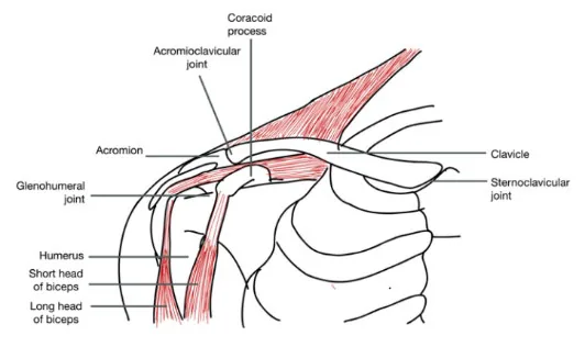
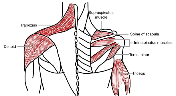
Why Check: Palpation can be very helpful to determine source of pain. Palpate for tenderness and swelling in a systematic manner, to assess for potential fractures and other abnormalities. Step-deformity, localized tenderness and swelling of the clavicle may indicate clavicular fracture. Tenderness at the biceps tendon has a Sensitivity of 57% and a Specificity of 72% for Bicipital Tendonitis.
Range of Motion (ROM)
AFMC level Pre-clerkship 3 · Clerkship 4
- Approach: Perform Active ROM, and then Passive ROM if Active ROM is limited. These can be integrated with examiner guiding the shoulder beyond patient’s active ROM.
- Active ROM:
- Screening Test – Apley Scratch Test: Have patient touch the tip of the opposite scapula (involves internal rotation and adduction) → Quick screen for shoulder ROM and subscapularis function
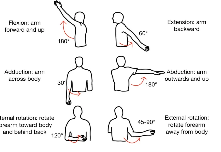
- Shoulder Flexion (Normal: 180°): Patient brings arms forwards all the way to the top
- Shoulder Extension (Normal: 60°): Patient moves arms backwards
- Shoulder Abduction (Normal: 180°):
- Painful Arc: Tends to occur between 60-120° during abduction; a positive painful arc mildly increases the probability of rotator cuff disease, while a negative painful arc moderately reduces the probability of rotator cuff disease (LR+ 3.7, LR- 0.36).
- Scapulothoracic Rhythm – <30°: Glenohumeral (GH) joint; 30-180°: 2:1 GH to Scapulothoracic joint
- If humerus and scapula move together as one complex → Suggests Adhesive Capsulitis
- Shoulder Adduction: Patient crosses arms in front of body
- Shoulder External Rotation (Normal: 45-90°): With arms relaxed at sides and elbows flexed to 90°, rotate forearms away from body.
- Shoulder Internal Rotation (Normal: Up to 120°): With arms relaxed at sides and elbows flexed to 90°, rotate forearms towards body and behind the back
- Passive ROM (perform if Active ROM limited):
- Crepitus: With hand over patient shoulder, move arm around to detect grinding/catching/cracking
- Adhesive Capsulitis: With passive ROM, this results in global restriction in all fields of movement.
Power Assessment
AFMC level Pre-clerkship 3 · Clerkship 4
- Standing Approach: With arms at side and elbows flexed to 90°
- Flexion: Clinician counter-supports placing one hand behind patient shoulder; with other hand, clinician pushes upper arm (just above elbow) posteriorly; patient resists motion.
- Extension: Clinician counter-supports placing one hand in front of patient shoulder; with other hand, clinician pushes upper arm (just above elbow) anteriorly; patient resists motion.
- External Rotation: Clinician stabilizes elbow with one hand; with other hand at wrist, push forearm inwards (medially); patient resists motion → Tests Infraspinatus and Teres Minor of Rotator Cuff
- Internal Rotation: Clinician stabilizes elbow with one hand; with other hand at wrist, push forearm outwards (laterally); patient resists motion → Tests Subscapularis of Rotator Cuff
- Sitting Approach: With arms abducted from sides at approximately 20°
- Abduction: Clinician hands just above patient elbows, and presses inwards; patient resists motion → Tests Supraspinatus of Rotator Cuff, as well as Deltoid and Trapezius muscles.
- Adduction: Clinician hands on inside of arms just above elbows, push outwards, patient resists motion.
- Reporting: For each muscle group tested, assess per the Medical Research Council Power Grading. If it is all 5/5, you can say “Power was 5/5 bilaterally in both upper and lower limbs”, and report specific deficits. NOTE: This scale is detailed in Chapter 31 – Screening Neurological Examination
Special Tests:
Rotator Cuff and Impingement Tests:
Drop Arm Test
AFMC level Pre-clerkship 3 · Clerkship 4
Say: I will assess the rotator cuff.
Do:
- Method 1: Patient abducts arms overhead, and actively brings arms back to side.
- Method 2: With patient arm abducted to 90 degrees, clinician “chops down” on arm.
- With either method 1 or 2, look for a sudden drop of the arm to one side.
Report: The drop arm test is negative.
Why Check: A positive drop arm test raises concern for a Complete Tear of Supraspinatus (most common site of rotator cuff tear). A negative drop arm test mildly reduces the probability of rotator cuff disease (LR- 0.82).
Painful Arc
AFMC level Pre-clerkship 3 · Clerkship 4
Say: I will continue to assess the rotator cuff. Please let me know if you feel any pain, and if so, where.
Do: Have patient actively abduct their arms, and look for signs of pain (typically between 60-120°) – commonly at lateral aspect or tip of the shoulder
Report: No signs of a painful arc on arm abduction.
Why Check: A positive painful arc mildly increases the probability of rotator cuff disease, while a negative painful arc moderately reduces the probability of rotator cuff disease (LR+ 3.7, LR- 0.36). A painful arc suggests External Subacromial Impingement (mechanical encroachment of bursa/rotator cuff tendons on subacromial space between humeral head and acromial arch)
Empty Can Test
Say: I will continue to assess the rotator cuff. Let me know if you feel pain, and if so, where.
Do:
- Abduct arm to 90° at the side then bring forward 30° with elbows extended and thumbs pointed down (“empty a can”)
- Clinician applies downward pressure on arms, patient actively resists.
- Look for signs of pain at tip of shoulder.
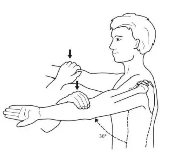
Report: No pain or weakness elicited – Empty Can Test is negative.
Why Check: The Empty Can test is used to assess for rotator cuff disease, where a positive test suggests External Subacromial Impingement (mechanical encroachment of bursa/rotator cuff tendons on subacromial space between humeral head and acromial arch). As per a JAMA Rational Clinical Exam article, the Empty Can test does not significantly alter the post-test probability of rotator cuff disease. A related test called the Full Can test (performed with arm in 45 degrees external rotation position with thumb pointed up instead of arm in full internal rotation with thumb pointed down) mildly increases the probability of rotator cuff disease (LR+ 2.4, LR- 0.37).
Lift Off Test
Say: I will continue to assess the rotator cuff.
Do: Have patient put their arm behind their back palm out at the level of the belt line. Have the patient push out against your hand, keeping the wrist straight. Look for signs of pain in the anterolateral shoulder or weakness.
Report: No pain or weakness elicited – negative Lift Off test.
Why Check: The Lift off test is used to assess for rotator cuff disease, specifically subscapularis tendon tears. A positive Lift off test mildly increases the probability of rotator cuff disease (LR+ 1.4-1.5, LR- 0.63-0.85).
Hawkins-Kennedy Test
Say: I will to assess the rotator cuff. Please let me know if you feel any pain, and if so, where.
Do:
- Shoulder flexed forward to 90°, elbow flexed 90°, with forearm parallel to floor
- Clinician passively rotates forearm clockwise to cause internal rotation at the shoulder.
- Look for signs of pain at shoulder.
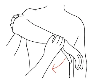
Report: No pain elicited – Hawkins-Kennedy Test is negative.
Why Check: A positive Hawkins Kennedy test mildly increases the probability of rotator cuff disease (LR+ 1.5, LR- 0.51); when Hawkins and Neer tests are both negative, this moderately decreases the probability of rotator cuff disease (LR- 0.43). A positive test suggests External Subacromial Impingement.
Neer’s Test
Say: I will continue to assess the rotator cuff. Please let me know if you feel any pain, and if so, where.
Do:
- Patient’s elbow in full extension; stabilize the shoulder by placing hand on posterior shoulder.
- Clinician passively lifts the arm to the ear (“Neer’s to the Ear”). Look for signs of pain. If pain is present, assess whether it is anterior or posterior shoulder pain.
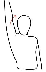
Report: No pain elicited – Neer’s Test is negative.
Why Check: Neer’s Test’s is used to assess for rotator cuff disease, where a positive test with anterior pain suggests External Subacromial Impingement (mechanical encroachment of bursa or rotator cuff tendons on subacromial space between humeral head and acromial arch), whereas a positive test with posterior pain suggests Internal Posterosuperior Glenoid Impingement (encroachment of rotator cuff tendons between humeral head and glenoid rim). When Hawkins and Neer tests are both negative, this moderately decreases the probability of rotator cuff disease (LR- 0.43).
Acromioclavicular (AC) Joint Tests:
Scarf Test
Say: I will assess the Acromioclavicular joint. Let me know if you feel pain, and if so, where.
Do:
- Patient places their hand on the opposite shoulder. Tell patient to allow hand to slide over shoulder during the test.
- Clinician applies a posterior force on the patient’s elbow in direction of the forearm to push the arm into the body while stabilizing the ipsilateral shoulder.
- Look for signs of pain in the area of the acromioclavicular joint.
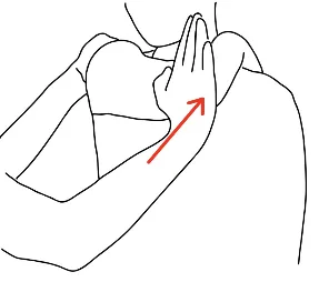
Report: No pain elicited around acromioclavicular joint – Scarf Test is negative.
Why Check: Pain elicited around acromioclavicular joint during Scarf’s test indicates acromioclavicular joint pathology, for example in Osteoarthritis (OA).
Labral Tests:
O’Brien’s Test
Say: I will assess the labrum. Let me know if you feel pain and if so, where.
Do:
- Patient in sitting or standing position with arm in 90 degrees of flexion and 10-15 degrees of adduction.
- Patient internally rotates shoulder so that thumb points to the floor.
- Clinician stabilizes the shoulder by placing hand posteriorly and with other hand applies downward force on lower arm as the patient resists with an upward force.
- The procedure is repeated with the shoulder in neutral position with thumb pointed to the ceiling.
- Look for pain or click deep in the shoulder joint.
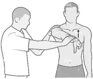
Report: No pain or clicking in either position – O’Brien’s test is negative.
Why Check: The O’Brien test assesses for tears of the labrum and is a two-part test. A positive test occurs when pain or clicking is produced deep in the shoulder with the first position but not in the second position. Superficial pain can indicate AC joint involvement. Studies show O’Brien’s test has a specificity ranging from 28-73% and a sensitivity ranging from 63-94%.
Stability Tests:
Sulcus Sign
Say: I will assess the shoulder for signs of instability.
Do:
- Both shoulders at rest. Clinician stabilizes contralateral shoulder and grasps arm just above elbow.
- Apply a downward pressure on the arm at the elbow. Look for Sulcus at the tip of shoulder.
Report: No sulcus seen on downward pressure of the arm – Sulcus Sign is negative.
Why Check: Sulcus Sign’s Diagnostic Value is in Ruling in Inferior Shoulder Instability, with a Sensitivity of 31% and a Specificity of 89%.
Anterior and Posterior Instability
AFMC level Pre-clerkship 1 · Clerkship 3
Say: I will continue to assess the shoulder for signs of instability.
Do:
- With both shoulders at rest position, clinician grasps one shoulder at the head of humerus and stabilizes by placing the other hand over the top of the shoulder.
- Clinician attempts to move the head of the humerus backwards and forwards in the glenohumeral joint (can be challenging if significant soft tissue present). Look for excessive movement.
- Note: Each shoulder should be assessed separately.
Report: No excess movement forwards or backwards to indicate instability anteriorly or posteriorly.
Why Check: Excessive movement forwards indicates anterior instability, while excessive movement backwards indicates posterior instability. Anterior shoulder dislocation can cause axillary neuropathy (axillary nerve is a branch of brachial plexus)
Biceps Tests:
Speed’s Test
Say: I will assess the biceps tendon. Let me know if you feel pain, and if so, where.
Do:
- Patient’s elbow extended, arm supinated and shoulder forward flexed at 45°.
- Clinician applies a downward pressure on the forearm. Look for signs of pain at the biceps tendon.
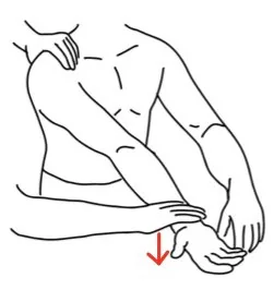
Report: No pain elicited at biceps tendon – Speed’s Test is negative.
Why Check: Speed’s Test’s Diagnostic Value is in Ruling Out Bicipital Tendonitis i.e. inflammation of long head of biceps tendon as it travels in intertubercular groove beneath transverse humeral ligament. Speed's test has a Sensitivity of 71% and a Specificity of 49% for bicipital tendonitis.
Yergason’s Test
Say: I will continue to assess the biceps tendon. Let me know if you feel pain, and if so, where.
Do:
- Patient’s arm at side, elbow flexed to 90° and hand in neutral position
- Grasp patient elbow, and forearm with other hand (just proximal to wrist)
- Attempt to pronate the forearm with patient resisting by trying to supinate.
- Look for signs of pain at the biceps tendon.
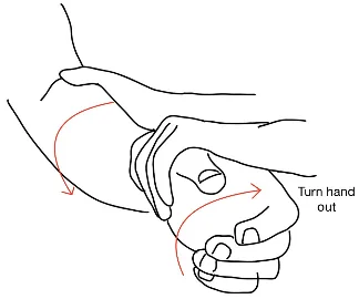
Report: No pain elicited at biceps tendon – Yergason’s Test is negative.
Why Check: A positive Yergason’s test is suggestive of Bicipital Tendonitis, along with Speed’s Test. Yergason's test has a Sensitivity of 64-68% and a Specificity of 30-61% for bicipital tendonitis.
Part 3: Select Special Tests – Not Part of Routine Shoulder Exam But Seen in Practice:
AC Joint Distraction Test
Say: I will continue to assess the Acromioclavicular Joint. Let me know if you feel pain, and if so, where.
Do:
- Patient adducts their arm behind their back, with dorsum of their hand resting on opposite buttock.
- Clinician stabilizes contralateral shoulder and applies additional adduction force (push on patient’s arm)
- Look for signs of pain in the area of the acromioclavicular joint.
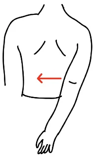
Report: No pain elicited around acromioclavicular joint – AC Joint Distraction Test is negative.
Why Check: Pain elicited around acromioclavicular joint during AC Joint Distraction test suggests acromioclavicular joint pathology, for example in Osteoarthritis (OA).
Apprehension Test, Anterior Relocation Test and Anterior Release Sign
Say: I will continue to assess the shoulder for signs of instability with moving the shoulder forward. Let me know if you feel any pain, and if so, where.
Do:
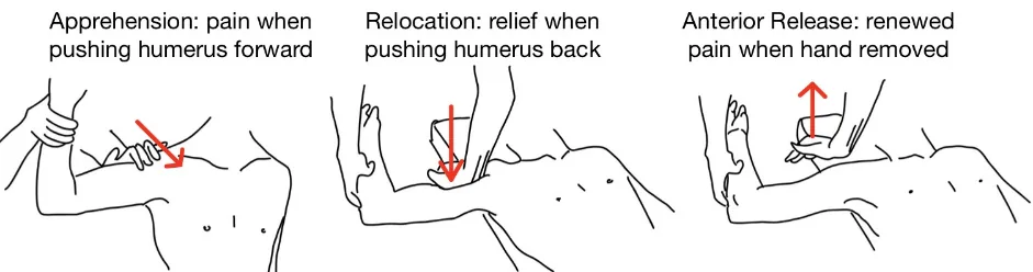
- While patient supine, passively abduct and externally rotate the arm at 90°.
- Apprehension Test: Clinician holds forearm with one hand, pulling humerus anteriorly with other hand. Look for signs of apprehension or pain (positive Apprehension Test).
- Relocation Test: Now reposition hand to push humerus posteriorly. Look for signs of relaxation or relief (positive Relocation Test).
- Anterior Release Sign: Now suddenly remove the hand that was pushing humerus posteriorly in relocation test. Look for signs of renewed apprehension or pain (positive anterior release sign).
Report: No signs of recurrent anterior subluxation of the shoulder with apprehension testing.
Why Check:
- The likelihood of anterior shoulder instability is moderately increased by positive anterior relocation test (LR+ 6.5, LR- 0.18) and positive anterior release sign (LR+ 8.3, LR- 0.09).
- Apprehension Test, Anterior Relocation Test, and Anterior Release Signs suggest recurrent anterior subluxation. Anterior Relocation test and Anterior release help rule in Anterior Shoulder Instability. Anterior shoulder dislocation can cause axillary neuropathy (axillary nerve – branch of brachial plexus).
- A positive apprehension test with anterior shoulder pain suggests External Subacromial Impingement (mechanical encroachment of bursa/rotator cuff tendons on subacromial space between humeral head and acromial arch).
- A positive apprehension test with posterior shoulder pain suggests Internal Posterosuperior Glenoid Impingement (encroachment of rotator cuff tendons between humeral head and glenoid rim).
A negative anterior relocation and release test suggests primary impingement (structural narrowing of subacromial space causing pain and dysfunction e.g. AC joint arthropathy, subacromial bursitis), while a positive anterior relocation and release test suggests secondary impingement (functional narrowing in specific positions re. excessive anterior translation of humeral head)
Helpful Videos to Review:
- Video 1: Shoulder Exam – McMaster MSK Examination Series (Link: SHOULDER EXAMINATION - McMASTER UNIVERSITY)
- Video 2: OSCE Shoulder Exam – Dr. Bob Lee Video (Link: https://www.youtube.com/watch?v=M1ilcE-y_NE)
- Video 3 and 4: Bates’ Visual Guide to the Physical Exam – Anatomy Review and Examining the Shoulders (Link: https://www.medportal.ca/ug/educational-resources/bates/lists/bates/musculoskeletal)
- Video 5 and 6: Dr. Janice Harvey’s Joint Assessment Made Easy – Shoulder Assessment Part 1 and 2 (Link: https://www.medportal.ca/ug/educational-resources/joint-assessment-made-easy)
Part 4: Relevant Likelihood Ratios – from Rational Clinical Examination JAMA series
Clinical Question 1: Does This Patient have Rotator Cuff Disease?2
| Procedure | Condition | LR+ (95% CI) | LR- (95% CI) |
|---|---|---|---|
| Pain Provocation Test – Painful Arc | Rotator Cuff Disease | 3.7 (1.9-7.0) (about +15%) | 0.36 (0.23-0.54) (about −30%) |
| Pain Provocation Test – Cross-Body Adduction | Rotator Cuff Disease | 1.9 (1.3-2.9) (about +15%) | 0.42 (0.26-0.68) (about −30%) |
| Pain Provocation Test – Hawkins | Rotator Cuff Disease | 1.5 (1.1-2.0) (about +15%) | 0.51 (0.39-0.66) (about −15%) |
| Pain Provocation Test – Neer | Rotator Cuff Disease | 0.98-1.6 (about −15%) | 0.60-1.1 (about −15%) |
| Pain Provocation Test – Yocum | Rotator Cuff Disease | 1.3 (0.75-2.3) (no change: 95% CI includes 1) | 0.53 (0.17-1.7) (no change: 95% CI includes 1) |
| Pain Provocation Test – Passive Abduction | Rotator Cuff Disease | 0.82 (0.58-1.1) (no change: 95% CI includes 1) | 2.6 (0.35-20) (no change: 95% CI includes 1) |
| Strength Tests – External rotation lag | Full Tear | 7.2 (1.7-31) (about +30%) | 0.57 (0.35-0.92) (about −15%) |
| Strength Tests – Internal rotation lag | Full Tear | 5.6 (2.6-12) (about +30%) | 0.04 (0.0-0.58) (about −45%) |
| Strength Tests – Drop Arm | Rotator Cuff Disease | 3.3 (1.0-11) (no change: 95% CI includes 1) | 0.82 (0.70-0.97) (about −15%) |
| Strength Tests – Dropping Sign | Full Tear | 3.2 (1.6-6.5) (about +15%) | 0.35 (0.15-0.83) (about −30%) |
| Strength Tests – Gerber (lift-off test) | Rotator Cuff Disease | 1.4-1.5 (about +15%) | 0.63-0.85 (about −15%) |
| Composite Test for Pain or Weakness – External rotation resistance | Rotator Cuff Disease | 2.6 (1.8-3.6) (about +15%) | 0.49 (0.33-0.72) (about −30%) |
| Composite Test for Pain or Weakness – Full can | Rotator Cuff Disease | 2.4 (1.5-3.8) (about +15%) | 0.37 (0.23-0.60) (about −30%) |
| Composite Test for Pain or Weakness – Patte | Rotator Cuff Disease | 1.4 (0.62-3.4) (no change: 95% CI includes 1) | 0.70 (0.34-1.5) (no change: 95% CI includes 1) |
| Composite Test for Pain or Weakness – Empty can (Jobe) | Rotator Cuff Disease | 1.3 (0.97-1.6) (no change: 95% CI includes 1) | 0.64 (0.33-1.3) (no change: 95% CI includes 1) |
| Composite Test for Pain or Weakness – Resisted abduction | Rotator Cuff Disease | 0.72 (0.55-8.1) (no change: 95% CI includes 1) | 2.1 (0.55-8.1) (no change: 95% CI includes 1) |
| Combination of findings – Hawkins and Neer (both positive) | Rotator Cuff Disease | 1.6 (0.87-2.8) (no change: 95% CI includes 1) | 0.43 (0.20-0.96) (about −30%) |
Clinical Question 2: Does this patient have Bicipital Tendonitis?3
| Procedure | Sensitivity | Specificity |
|---|---|---|
| Speed’s Test | 0.71 | 0.49 |
| Palpation for Tenderness | 0.57 | 0.72 |
| Yergason’s Test | 0.64-0.68 | 0.30-0.61 |
Clinical Question 3: Does this patient have Inferior Shoulder Instability?4
| Procedure | Sensitivity | Specificity |
|---|---|---|
| Sulcus Sign | 0.31 | 0.89 |
Clinical Question 4: Does this patient have Anterior Shoulder Instability?56
| Procedure | LR+ (95% CI) | LR- (95% CI) |
|---|---|---|
| Anterior Release Sign | 8.3 (3.6-19) (about +30%) | 0.09 (0.03-0.27) (about −45%) |
| Anterior Relocation Test | 6.5 (3.0-14) (about +30%) | 0.18 (0.07-0.45) (about −30%) |
References
-
Recommendations. Choosing Wisely Canada. Accessed July 22, 2022. https://choosingwiselycanada.org/recommendations/#list ↩
-
Hermans J, et al. Does this patient with shoulder pain have rotator cuff disease? JAMA. 2013;310(8):837-847. ↩
-
Chen HS, et al. A comparison of physical examinations with musculoskeletal ultrasound in the diagnosis of biceps long head tendinitis. Ultrasound Med Biol. 2011;37(9):1392-8. ↩
-
T’Jonck L, et al. The relationship between clinical shoulder tests and the findings in arthroscopic examination. Genes Sport. 2001;34:15-24. ↩
-
Barth JR, et al. The bear-hug test: a new and sensitive test for diagnosing a subscapularis tear. Arthroscopy. 2006;22(10):1076-1084. ↩
-
Gross ML, Distefano MC. Anterior release test. Clin Orthop. 1997;339:105-108. ↩