Medical Foundations 3
Chapter 17Select Topics in Pediatric Growth and Development
Introduction
- In pre-clerkship clinical skills, you will discuss an approach to the Newborn History (Session 8) and the Adolescent History (Session 9). The newborn exam is included as a reference for clerkship, and is a focus of the Pediatrics clerkship. This Chapter will cover core topics for the Newborn History, which has connections to the Obstetrical History (Chapter 20).
- Clinical Skills Videos for the Newborn Exam:
- Bates’ Visual Guide to the Physical Exam: (Requires Medportal Login)
- JoVE Clinical Skills Videos – The Comprehensive Newborn Exam: (Create account via mcmaster.ca email)
Part 1: Focused History – Based on Chief Complaint (Begin with Open-Ended Questions)
Newborn Visit Info
Antenatal History
- Maternal Health History:
- Pre-existing medical conditions
- Maternal medications
- Maternal substance use (smoking, EtOH, marijuana, IVDU)
- Maternal blood type; Hx of previous child w/ ABO or Rh incompatibility
- Obstetrical History & Current Pregnancy:
- GTPAL (G=gravidity; T=term; P=preterm; A=abortions; L=living); alternatively: GP status (G= gravidity; P=parity)
- Past pregnancies: Year, sex, weight, GA, preterm, premature rupture of membranes, need for induction, length of labour, method of delivery (if C-section, ask why), complications
- If abortion: Ask if instruments involved (i.e. Dilation and Curettage, D&C)
- Document previous ectopic & molar pregnancies
- Maternal serologies during this pregnancy (e.g. rubella, HIV, syphilis, hepatitis B)
- Vaccinations during this pregnancy (e.g. Tdap)
- Previous complications: Gestational diabetes (GDM), hypertensive disorders of pregnancy (gestational hypertension, pre-eclampsia, etc.), antepartum hemorrhage/bleeding, etc.
- Antenatal ultrasounds: Last ultrasound? BPP? Estimated fetal weight? Any abnormalities (e.g. anatomic, abnormal amniotic fluid volumes, intrauterine growth restriction, etc.)?
- GTPAL (G=gravidity; T=term; P=preterm; A=abortions; L=living); alternatively: GP status (G= gravidity; P=parity)
- Prenatal Screening:
- Were any tests done? If so, which ones? (e.g. First Trimester Screen, Quad Screening, Integrated Prenatal Screening, Non-Invasive Prenatal Screening) (Note: Clinicians only contacted with results if positive)
- Did tests require follow-up (e.g. amniocentesis, chorionic villus sampling)? What were results?
Labour & Delivery (L&D)
4 essential questions to always ask at L&D triage
- Contractions/labour: When did they start? How often (X minutes)? How long do they last? Getting bigger or closer together?
- Fluid/rupture of membranes: Any water breaking? When? Big gush or small trickle? What colour (clear vs. green vs. red)?
- Bleeding: When did it start? How many pads? What colour (red vs. brown vs. pink)? Any pain? Any trauma, intercourse or instrumentation? Last ultrasound + placenta location?
- Fetal movements: Still feeling kicks? Any change to FM? Kick count okay (≥6 in 2h)?
Anticipating the need to resuscitate a newborn as a result of fetal distress increases the likelihood of a successful resuscitation. 4 pre-birth questions to identify newborns ↑ risk of resuscitation are:
- What is the gestational age? (<36 weeks or >40wks + 6d ↑ risk)
- Was amniotic fluid clear? (meconium-stained amniotic fluid – may indicate fetal distress, ↑ risk)
- Multi-gestations? (↑ risk; also informs appropriate equipment amounts to have at-the-ready)
- Any additional risk factors? (↑ risk of needing neonatal resuscitation):
- Antepartum: No prenatal care; Hypertensive disorders in pregnancy (e.g. GHTN, Pre-eclampsia or eclampsia); Oligohydramnios; Polyhydramnios; Intrauterine Growth Restriction (IUGR); Significant fetal malformations or anomalies; Fetal anemia; Fetal hydrops; Fetal macrosomia
- Intrapartum: Emergency C/S; Forceps or Vacuum-Assisted delivery; Breech/abnormal presentation; Atypical/Abnormal Fetal Heart Rate Patterns; Maternal general anesthesia; maternal magnesium therapy (e.g. for eclampsia); placental abruption; intrapartum bleeding; chorioamnionitis; narcotics administered to mother within 4 hours of delivery; shoulder dystocia; meconium-stained amniotic fluid; or prolapsed umbilical cord
- Number of Weeks Gestation: Pre-term <37 wk; Early term: 37wk – 38wk + 6d; Full Term: 39wk – 40wk + 6d; Late Term: 41wk – 41wk + 6d; Post-Term ≥42 wk 0d
- GBS status → pregnant mothers tested at 35 weeks (result valid for 5 weeks)
- For pregnancy at term (≥ 37 weeks gestation) with positive GBS or unknown GBS status AND Prolonged ROM (> 18 hours) treated with IV Penicillin G.
- For preterm prelabour rupture of membranes (PPROM – < 37 weeks gestation) with positive GBS or unknown GBS status, treated with IV Penicillin G.
- Adequate Antibiotic Prophylaxis: ≥1 dose of IV Penicillin G > 4 hours before delivery
- Rupture of membranes (ROM): Usually spontaneous (<1 hour between ROM and start of contractions in a term fetus ≥37 weeks gestation). Artificial ROM accelerates labour.
- Prelabour rupture of membranes (PROM): ROM ≥ 1 hour before onset of contractions in a term fetus ≥37 weeks gestation
- Prolonged ROM: (>18 hours) → ↑ infection risk → may treat prophylactically with antibiotics
- Labour:
- Induction of Labour: Helping patient to go into labour e.g. with cervical ripening interventions (e.g. prostaglandins, balloon dilatation of cervix for an unfavourable cervix) or oxytocin or artificial rupture of membranes for a favourable cervix.
- Augmentation of Labour: help ripen the cervix for labour (e.g. prostaglandins); Augmentation agents help with contractions (e.g. oxytocin).
- Method of Delivery (Spontaneous vaginal, Vaginal with instrumentation, C-Section)
- Instrumentation: vacuum-assisted, forceps-assisted
- C-Section: elective, urgent, emergent (if so, what is the indication?)
- Sepsis Risk Factors: Chorioamnionitis, Maternal intrapartum T >38°C, ROM duration >18hrs; delivery < 37 weeks’ gestation
- Routine Newborn Medications
- Vitamin K: Prevents hemorrhagic disease of newborn
- Hepatitis B Vaccine (if indicated)
APGAR Scores
AFMC level Pre-clerkship 1 · Clerkship 4
- APGAR Scores at 1 and 5 minutes: Appearance, Pulse, Grimace, Activity, Respiration.
- Significance: Low APGAR scores alone do not predict poor neurologic outcome; rather, persistent low scores at 10, 15 and 20 minutes ↑ risk of poor outcomes. A change in APGAR scores over time is a useful indication of resuscitation response.
- Evidence: There is poor inter-observer reliability of APGAR scores, especially in very unwell infants1
Neonatal Resuscitation
- Did clinical team request additional support at delivery? What was done?
Neonatal Course
- Length of stay in hospital/age at time of discharge
- NICU admission: indication, length of admission
- Jaundice: Did they need to go under the UV lights (phototherapy?) If so, for how long?
Newborn Screening (NBS):
- Heel-prick test (Publicly funded): includes inborn errors or metabolism, endocrine disorders (congenital hypothyroidism, congenital adrenal hyperplasia), Sickle Cell, CF, and Severe Combined Immune Deficiency (SCID).
- Results only communicated if positive. For more information, visit www.newbornscreening.on.ca
- Critical congenital heart disease screening: Pre- and Post-ductal oxygen saturation test (probe on right hand/wrist and one of the feet) – see Physical Exam below
Audiology Testing (component of Newborn Screening)
AFMC level Pre-clerkship 1 · Clerkship 1
- Screening Tests (Performed at Any Age): Evoked optoacoustic emissions; automated auditory brainstem response (ABR) – publicly funded test for permanent hearing loss
- Diagnostic Tests: Automated ABR (3-6 months); Visual Reinforced Audiometry (6-24 months); Play Audiometry (2-4 years); Conventional Pure Tone Audiometry (4+ years)
- Why Check: Abnormal hearing screens require further workup; parents identify subtle hearing loss up to 12 months earlier than physicians identifying it. Thus, if parents have any concerns with hearing in their child, audiometry should be undertaken.2
Growth
- Birth parameters: Weight, length, head circumference
- Birth Weight: Macrosomia (>4500g); Average term neonate (2500-4500g); Low Birth Weight (1500-2500g); Very Low Birth Weight (1000-1500g); Extremely Low Birth Weight (<1000g)
- Discharge parameters: Weight, length, head circumference
- Current parameters: Weight, length, head circumference at time of assessment
- Expect weight loss after birth, but should not exceed 10% loss from birth weight; should regain birth weight within 10-14 days after birth
Feeding/Intake
- WHO Recommendation: Early initiation of breastfeeding within 1 hour of birth; exclusive breastfeeding for the first 6 months of life; and introduction of nutritionally adequate and safe complementary (solid) foods at 6 months together with continued breast feeding up to 2 years of age or beyond.
- Breastfeeding
- How often does the infant feed (expect q2-3h, or 8-12 times in 24 hours)?
- For how long does the infant feed at each breast (expect 15-30min per breast)?
- Has the infant been able to latch well?
- Does your infant become very sweaty (diaphoretic), fatigued, tachypneic, or cyanotic during feedings? – Screen for Congenital Heart Defects
- Reflux/emesis: quantity, frequency, appearance (blood/bile?)
- Have you had consultation with a lactation specialist?
- Supply: let-down and emptying? Pumping between feeds?
- Formula: What formula? How is it prepared? What is volume per feed, and how many feeds/day?
- Other feeds: New foods introduced? When? (Typically exclusive BF/formula until 6 months old)
- Helpful resource to discuss infant feeding: Informed Decision Making: Having Meaningful Conversations Regarding Infant Feeding3
Output
- Stool
- Colour: If breastfed, expect transition ~ day 4-5 from meconium (black) → mustard (yellow) stool, runny, may have small whitish curds. If formula-fed, light brown/greenish with consistency of peanut butter.
- Frequency: Number of stools/diapers per day?
- Passage of meconium within first 24h of life? – Screen for intestinal obstruction/atresia, Cystic Fibrosis (meconium ileus)
- Urine
- In first 4 days of life, expected number of wet diapers equal to day of life (1 wet diaper day 1, 2 wet diapers day 2, etc.). By day 5 going forward, expect 6-8 wet diapers per day.
- Colour: Assess for Cysteine crystals (dehydration) vs hematuria
- Quantity/frequency: Number of wet diapers/day (<3/24h→ concern re. dehydration)
Activity/Sleep
- Activity level/energy: is infant awake, alert, responsive? — Red flags: irritability, lethargy
- How often does your infant sleep? – Expect 16-22 hrs/day
Address Common Parent Concerns
- Congestion: Expect first 4-6 weeks – try saline drops in nose/nasal suction prior to feeds.
- Unilateral or Bilateral Eye Discharge: Can be normal due to obstructed lacrimal ducts, should clear up with time (Not normal if eye is red – may suggest bacterial vs. viral conjunctivitis)
- Swollen Breasts: Hormonal contribution of mother, should dissipate with time
Failure to Thrive (FTT)
- What is Failure to Thrive?
- Variable definitions – Growth rate less than expected for a child, particularly concerning when <3rd percentile and/or crossing 2 major percentile lines over time.
- Inorganic Etiologies of FTT (Most Common Causes):
- Food: Formula preparation, feeding techniques
- Parental: Child abuse & neglect; maternal depression; mental health considerations; substance abuse
- Family: Violence; marital discord
- Society: Low SES, poor social support networks
- Organic Etiologies of FTT:
- Cardiac: Congenital Heart Disease (diaphoretic with feeding)
- Pulmonary: Bronchopulmonary Dysplasia (BPD – may be seen in premature infants); Cystic Fibrosis
- Hematology: Iron-deficiency Anemia (e.g. Excess cow’s milk intake); Malignancy
- GI – Motility: Craniofacial pathology; tracheoesophageal fistula; GERD
- GI – Obstruction: Pyloric Stenosis (immediate post-prandial, non-bilious, forceful vomiting); Malrotation/Volvulus (twisting of bowel around superior mesenteric artery, bilious vomiting); Hirschsprung’s Disease (aganglionic megacolon)
- GI – Absorption: Diarrhea; Inflammatory Bowel Disease; Cystic Fibrosis; Celiac Disease
- Endocrine: Hypothyroid; Rickets
- Metabolic & Congenital: Fetal Alcohol Syndrome; Inborn Errors of Metabolism
- Infectious: Tuberculosis, HIV, Hepatitis
- Neurological: Hydrocephalus; Intracranial Tumors; Muscle Weakness
Subsequent Visits
Milestones
note that milestone charts differ in milestone’s associated age, e.g. age at which 50% are expected to reach it, vs. 75% is when milestone is listed.
Denver II – [1, 2] Rourke Baby Record – [1, 3]
- Why Check: A critical part of well-child visits in community practice, as well as any time you examine a child.
- Resource for Parents & Clerks – Nipissing District Developmental Screen (NDDS): Includes several useful resources for parents and Health-care providers
- Alternate Resource for Clerks – Med-U.org Developmental Milestones (https://med.stanford.edu/content/dam/sm/pediatricsclerkship/documents/5-Developmental-Milestones-MedU.pdf): Used in Pediatric Clerkship.
- Denver Developmental Screening Test (Denver II): A standardized test screening for developmental delay combining a parental report of child’s skills in major developmental areas with examiner observations. Children with abnormal scores are at higher risk of poor school performance (26% vs. 10% with normal screens), and lower IQ (<89 in 43% of children with abnormal Denver II). It also has a higher incidence rate in children with low SES backgrounds.4
- Recording Well Child Visits – Rourke Baby Record (https://www.rourkebabyrecord.ca/downloads): Evidence based structured interview form used in primary care for well-child visits from 0-5 years; structured to screen for concerns in growth, nutrition, development, behavior, immunizations, and abnormal physical findings or congenital anomalies. It highlights areas of focus for each visit with evidence-based maneuvers but is not meant to replace physician clinical acumen on what to include as part of each visit.5
Family-Centred Weight Discussion
- Sample Questions to Begin Conversation with Pregnant Patients about Weight (Note: Depending on the developmental stage of the patient, consider speaking with parents alone)
- Would it be alright if we discussed your child’s weight?
- Are you concerned about your child’s health?
- Are you concerned about your child’s weight?
- *Source: 5As of Pediatric Obesity Management. Canadian Obesity Network. www.obesitynetwork.ca
Approach to Childhood Disability6
- F Words:
- Traditional Medical Model Word – Fixing: Acute care medicine approach
- New Word 1 – Function: encourage the development and practice of function without regard to how nicely it is achieved.
- New Word 2 – Family: Focus on supports and resources for families to help make informed decisions.
- New Word 3 – Fitness: Emphasis on health-promoting orientation, not simply focusing on remediation of their ‘disabilities’.
- New Word 4 – Fun: First, find out what they want to do; then adapt as needed to allow children to pursue these activities/goals. Do not worry about expecting children to do things “normally”. It is the doing, rather than a superior level of accomplishment, that is most meaningful to most children.
- New Word 5 – Friends: Quality of relationships, not the number, that is important.
- New Word 6 – Future: All children, including disabled children, are in a constant state of “becoming”. Think about the future in a positive way, right from the start. We can ask parents and children with disabilities, at any time, about their expectations and dream for a future that is possible – and not decide for them what is impossible.
Publicly Funded Vaccination Schedules
- Vaccinations and Well-Child Visits: In community practice, well-child visits tend to be organized around the Publicly Funded Vaccination Schedule. Given the gap between 18-month vaccines and 4-year vaccines, therefore the 18-month visit is both important and thorough (e.g. Developmental screening with the LookSee 18-month checklist by NDDS; Autism Screening such as M-CHAT if symptoms of social difficulty/autism).
- Recent Change: As of April 2016, the Ontario government is now also publicly funding and adding to the Vaccination schedule the HPV Vaccine for all genders
- Current Ontario Publicly Funded Vaccination Schedule: https://www.ontario.ca/page/ontarios-routine-immunization-schedule.
- National Vaccination Schedule by Province/Territory: https://www.canada.ca/en/public-health/services/provincial-territorial-immunization-information/provincial-territorial-routine-vaccination-programs-infants-children.html.
SSHADESS: Adolescent Interview
- Purpose of Targeted SSHADESS Interview: Complete psychosocial context to screen for risky behaviors that may result in top 3 causes of adolescent death – (1) Accidents, (2) Homicide and (3) Suicide
- Confidentiality Statement: As with all of our visits, the information we discuss will be kept confidential. I might take notes and put those in your record here, but everything will be stored securely. There are some instances where I may be required by law to break confidentiality, for example if I believe there is immediate risk of harm to yourself or others. Does this sound okay with you?
- Components of SSHADESS Interview:
- Strengths: What do you like to do/what are you good at?
- School: Grade and name of school, academic performance; Special programs, recent changes in school, suspensions/expulsions? Plans for future education? Any bullying?
- Home: Who lives with you? What are relationships like at home? Any new people in home environment? Have the police or CAS ever been involved with your family? (see Social History for information on CAS/FACS and physicians’ duty to report). Why did CAS get involved? Are they still involved?
- Activities: What do you like to do for fun? What do you do outside of school (e.g. extracurriculars, sports, jobs/employment)?
- Drugs/substance use: Do any of your friends smoke (cigarettes, vaping, marijuana, etc.) use alcohol or other drugs? Have you ever tried these things? How much do you use and how often? What situations do you use those substances in? Do your family members use drugs, including alcohol and tobacco? Do you use any performance enhancing drugs?
- Eating/Emotions: Describe what a typical day of eating looks like for you. Do you have any concerns about your body weight, shape, or size?
- Mood screen: depression, mania, anxiety (e.g. Tell me about what worries you).
- Screen for suicidality and self-harm: Have you ever thought about hurting yourself or someone else? Have you ever hurt yourself? Have you ever thought life is not worth living?
- Sexuality: What is your name? What are your pronouns? Have you ever been in a relationship? Who are you attracted to? What kind of sex have you engaged in? How many partners have you had? Do you use contraception and/or protection? Have you ever been pregnant? Do you have any symptoms or concerns about STIs?
- Safety: Seat belt use in car; driving safety; violence at home; physical, emotional, or sexual abuse; ridden in a car with a driver who is drunk or high?
Past Medical History (PMHx)
- Any conditions diagnosed based on pre-natal screening (e.g. Down’s Syndrome, etc.)
- Any neonatal complications or any other diagnoses?
Past Surgical History (PS Hx)
- Neonatal procedures (e.g. tongue tie release, circumcision) and surgeries (e.g. abdo surgery for bowel malrotation)
Family Medical History (FMHx)
- Review any maternal and paternal family history, current medical conditions, etc.
- Screening for genetic/metabolic disorders:
- Consanguinity
- Sudden/unexpected infant deaths or deaths in childhood
- Recurrent miscarriages/stillbirths
- Genetic disorders (e.g. Down Syndrome)
- Developmental delays or developmental disorders
- Congenital anomalies (e.g. heart defects, limb abnormalities, dysmorphisms)
Social Hx
- Parents: Smoking, Alcohol Intake (↑ Alcohol intake associated with ↑ breast cancer risk in females), Illicit Drug use, Nutrition, Family & Social Support Network
- Previous CAS/FACS and/or police involvement with the family (see below re. duty to report to CAS/FACS)
Medications Hx & Allergies
- Vitamin D (for any infant exclusively/partially breastfed) – 400 IU/day, or 800 IU/day in Northern Canada (>55th latitude)
- Iron supplementation (for most premature neonates)
Part 2: General Physical Exam Maneuvers in Well-Child Visits – Vitals and Measures of Growth
Pediatric vitals
RR – [1, 3] BP – [2, 4]
Note: Pediatric Vital Normal Values depend on age of the child – from PALS (American Heart Association)
Heart Rate (HR) – beats per minute (bpm)
| Age | Awake HR | Sleeping HR |
|---|---|---|
| Neonate | 100-205 | 90-160 |
| Infant | 100-180 | 90-160 |
| Toddler | 98-140 | 80-120 |
| Preschooler | 80-120 | 65-100 |
| School-Age Child | 75-118 | 58-90 |
| Adolescent | 60-100 | 50-90 |
Respiratory Rate (RR) - breaths per minute
| Age | RR |
|---|---|
| Infant | 30-53 |
| Toddler | 22-37 |
| Preschooler | 20-28 |
| School-Age Child | 18-25 |
| Adolescent | 12-20 |
| Age | Systolic BP (mmHg) | Diastolic BP (mmHg) | Mean Arterial Pressure (MAP) |
|---|---|---|---|
| Birth (12hrs, <1000g) | 39 – 59 | 16 – 36 | 28 – 42 mmHg |
| Birth (12hrs, 3kg) | 60 – 76 | 31 – 45 | 48 – 57 mmHg |
| Neonate (96 hrs) | 67 – 84 | 35 – 53 | 45 – 60 mmHg |
| Infant (1-12mo) | 72 – 104 | 37 – 56 | 50 – 62 mmHg |
| Toddler (1-2yo) | 86 – 106 | 42 – 63 | 49 – 62 mmHg |
| Preschooler (3-5yo) | 89 – 112 | 46 – 72 | 58 – 69 mmHg |
| School-Age Child (6-9yo) | 97 – 115 | 57 – 76 | 66 – 72 mmHg |
| Pre-Adolescent (10-11yo) | 102 – 120 | 61 – 80 | 71 – 79 mmHg |
| Adolescent (12-15yo) | 110 – 131 | 64 – 83 | 73 – 84 mmHg |
| Temperature | Rectal (gold standard) | Oral | Tympanic | Axillary |
|---|---|---|---|---|
| Normal (°C) | 36.6-38 | 35.5-37.5 | 35.8-38 | 36.5-37.5 |
Why Check:
- Respiratory Rate: RR>50 (2-12 months) or RR>40 (>12 months) is suggestive of pneumonia. This test has a sensitivity of 40% (95%CI 33-46), and a specificity of 74% (95% CI 64-69)14
- Blood Pressure: Measurement by auscultation in child’s right arm in seated position is preferred method. Screen BP measurements in children >3 years old, unless risk factors present. Hypertension in children is a SBP or DBP >95th percentile measured on 3 occasions and standardized to reference charts for gender, age & height. If a child has HTN, it is more likely to be from a secondary cause, and a workup should be done.15
- Vital Signs indicating need for rapid assessment and potential cardiopulmonary support (per PALS):
- Irregular respirations or RR > 60 breaths per minute
- HR for Child ≤ 2 years of age: <80 bpm or >180 bpm
- HR for child > 2 years of age: <60 bpm or >160 bpm
- Fever with Petechiae
Pediatric Growth Curves
AFMC level Pre-clerkship 2 · Clerkship 3
Say: I will begin with measuring the child’s height, weight and head circumference. Before we begin, could we have your child change so they are only in their diaper?
Setup: Requires a weight scale, a method of measuring height, and a tape measure.
Do: Measure patient’s weight using appropriate scales. Measure height, ensuring to extend Lower Limbs as much as possible. Measure Head Circumference using firm measuring tape around widest possible circumference of head – above eyebrows and ears, in a transverse plane. Input data into WHO Growth Chart Centiles. Compare current centiles with past centile measurements (if available).
Report: The measurements today show your child is at 50th percentile for height and 50th percentile for weight.
Why Check:
- Interpret in the Context of the Patient, Focus on Healthy Habits: When plotting Height, Weight & Head Circumference on the patient’s Growth Charts, interpret the resulting growth curves in the context of the patient –concepts of normal for you, and in balance weight and height with emphasis on healthy eating habits and active participation in physical activity are important.
- Newborns in the First 2 Weeks: Newborns can lose up to 10% of their birth weight upon discharge and be considered within the range of normal – they also typically lose a small amount of weight once discharged home, typically bottoming out around Day 4-5 before beginning to gain weight. If breast fed, important to ask if stools have begun to transition to mustard yellow stools, as this will tend to occur in Day 4-5 after meconium has passed, and is evidence that breast milk is being taken in.
- Premature Infants and Growth Charts: Shift growth curve left based on 40 – # weeks gestation.
- Expected Adult Height: Male: Avg parental ht +2.5” (6.35cm); Female: Avg parental ht –2.5” (6.35cm).
- The following are rules of thumb for expected rates of growth:
| Birth-3 months | 3 – 6 months | 6 – 12 months | 1 -2 years | 2 years – adolescence | |
|---|---|---|---|---|---|
| Weight | 25-30 g/day Regain BW @ 2 weeks | 20 g/day Double BW @ 4-6 mo | 10 g/day Triple BW @ 12mo | 250 g/month | 2.3 kg/year |
| Height | 25 cm/year – 50% Length ↑ at 12 months | 25 cm/year – 50% Length ↑ at 12 months | 25 cm/year – 50% Length ↑ at 12 months | 12.5cm/year | 6.25cm/year |
- Question – What Growth Curves Do I Use to Chart? Recommendation is to use WHO growth curves in first 24 months of life, after which you may continue using WHO growth curves, or switch to use of CDC curves. This recommendation is based off differences in the methodology behind each curve’s construction.16
- WHO Growth Charts: Developed with longitudinal data of exclusively breastfed infants across cultures, describing healthy growth under optimal conditions, with no ethnic group variation.
- CDC Growth Curves: Describe a particular cohort of children’s growth in USA over 30-year span
- Pediatric Obesity: Obesity status in children defined using BMI age- and gender-specific growth charts. CDC defines pediatric obesity as >95th percentile; WHO defines pediatric obesity as >97th percentile.
- Edmonton Obesity Staging System – Pediatrics (EOSS-P)17: Similar to EOSS used for Adults, but focuses on the 4 M’s: Metabolic, Mechanical, Mental, and Milieu:
| EOSS-P Stage | Metabolic | Mechanical | Mental | Milieu |
|---|---|---|---|---|
| Stage 0 | None | None | None | None |
| Stage 1 | Mild (IGT, Pre-HTN, Mild Lipid abnormalities, mild fatty infiltration of liver/↑ transaminases) | Mild (OSA not requiring PAP therapy, Mild MSK pain not interfering with ADLs, GERD) | Mild (ADHD, LD, Mild body image pre-occupation, occasional emotional/binge-eating, bullying, mild develop. delay) | Mild (Minor relationship problems, minor limitations in caregivers’ ability to support child’s needs) |
| Stage 2 | Moderate requiring pharmacotherapy (T2DM, HTN, DLD, PCOS, Mod-Severe Fatty infiltration of liver) | Moderate (OSA requiring PAP therapy, GERD, MSK pain limit activity, moderate ADL limitation) | Moderate (Major depression, Anxiety, frequent Binging, Significant body image disturbance, mod. Develop. delay) | Moderate (Moderate relationship problems, significant bullying, significant limitations in caregivers’ ability to support child’s needs) |
| Stage 3 | Uncontrolled metabolic complications (T2DM with complications, uncontrolled HTN, FSGS, marked lipid abn, Marked ↑ Liver Enz/ dysfunction, gall stones) | Severe (OSA requiring PAP therapy + O2, limited mobility, Dyspnea sitting/sleeping) | Uncontrolled (School refusal, daily binge eating, severe body image disturbance) | Severe (Severe relationship problems, caregivers unable to support child’s need including family violence, dangerous environment (home/neighborhood/school) |
Developmental Milestones18
AFMC level Pre-clerkship 1 · Clerkship 2
| Age | Gross Motor | Fine Motor | Communication/Social | Cognitive/Adaptive |
|---|---|---|---|---|
| 2 mo. | Lifts head/chest when prone | Eyes track past midline | Alerts to sound Social (reciprocal) smile | Recognizes parent |
| 4 mo. | Rolls front to back | Grasps a rattle | Laughs; Soothed by parent’s voice | Orients head to direction of a voice |
| 6 mo. | Sits with little to no support | Reaches with 1 hand Transfers objects | Babbles. Developing stranger anxiety | Feeds self |
| 9 mo. | Pulls to stand | Developing; Immature pincer grasp; Bangs two objects together | Says mama/dada indiscriminately Waves bye-bye | Plays gesture games |
| 12 mo. | Stands/walks alone | Fine pincer grasp | One word other than mama/dada; Follow 1-step commands with gesture | Points to a desired object |
| 15 mo. | Stoops and recovers | Scribbles in imitation | Uses 3-5 words | Uses spoon & cup Turns pages in a book |
| 18 mo. | Runs well | Builds a tower of 3 cubes | Points to 1-3 body parts 15-20 words | “Helps” in the house |
| 24 mo. | Throws ball overhand Kicks a ball | Copies a line with a crayon | Speaks in 2-word combinations 50+ word vocabulary 50% language intelligible to a stranger; Parallel play | Removes an article of clothing |
| 36 mo. | Pedals a tricycle | Copies a circle | Speaks in 3-word sentences 250 words; 75% language intelligible to a stranger | Brushes teeth with help |
| 48 mo. | Hops | Copies a square or cross | >250 words; 100% language intelligible to a stranger; Plays cooperatively with a group | Knows 4 colours |
| 60 mo. | Skips | Copies a triangle | Defines simple words Uses 5-word sentences | Dresses self |
Why Check: Domains of development include Gross motor, fine motor, communication/social, and cognitive/adaptive, where milestone charts outline developmental progression over time. Milestone charts (example above) outline developmental progression over time. It is recommended that milestone ages listed in milestone charts should be based on the oldest age by which the skill should have been achieved19. Red flags for Developmental Milestones include:
- Gross Motor: No head control by 3-4 months; No independent sitting by 8-9 months; No independent steps by 18mo-24mo
- Fine Motor: Fisting >50% of time at 4 months; Not reaching for objects by 6 months; Not transferring objects by 8 months; Poorly developed pincer grasp at 15 months; Hand dominance earlier than 18 months
- Language: No babbling by 10-12 months; No attempt at words by 18 months; <25-50 words at 24 months
Developmental surveillance involves ongoing monitoring of development, identification of risk factors and elicitation of parental concern. Clinicians should remain vigilant to deficits in children’s performance across developmental domains and consider further evaluation for children whose development does not meet age-expected milestones19. Developmental surveillance is part of the Rourke Baby Record, where absence of any item suggests considering further assessment of development5. The Canadian Task Force on Preventive Healthcare recommends against screening for developmental delay using standardized tools in children aged 1 to 4 years with no apparent signs of developmental delay and whose parents and clinicians have no concerns about development19. In Ontario, an exception is the enhanced 18-month well-baby visit, where clinicians perform developmental screening using the LookSee 18 Month Checklist by NDDS.
Part 3: Focus on the Newborn Exam (Not discussed in preclerkship clinical skills– included for reference)20212223
- Key Idea: If you’re concerned about a newborn, have a low threshold to call for help – patient safety comes FIRST.
- The newborn exam facilitates early identification of anomalies and health problems requiring immediate intervention. If one anatomic malformation is found, the newborn should be evaluated for associated abnormalities. The exams listed are organized according to a Head-to-Toe Approach.
- Pro Comp Connection: Depending on setting, the exam room may be different from the room where parents and baby are staying; parents may have also been discharged, with baby kept for monitoring. Where possible, consider offering parents the opportunity to observe the newborn exam, and to talk through your findings with them including reassuring findings.
Notes on Vitals, APGAR Scores, Growth Parameters, and Pre- vs. Post-Ductal Oxygen Saturation Screen
Newborn vitals24
RR – [1, 3] BP – [2, 4]
- Temperature: 36.5-37.5°C (Axillary temperature) - measure in an open crib
- RR: 40-60 breaths per minute → count over a full minute
- HR: Note that HR may decrease to 90 bpm with sleep; an increase of HR with stimulation is reassuring
Heart Rate (HR) – beats per minute (bpm)
| Age | Awake HR | Sleeping HR |
|---|---|---|
| Neonate | 100-205 | 90-160 |
- BP: Use an appropriate neonatal-sized BP cuff
| Age | Systolic BP (mmHg) | Diastolic BP (mmHg) | Mean Arterial Pressure (MAP) |
|---|---|---|---|
| Birth (12hrs, <1000g) | 39 – 59 | 16 – 36 | 28 – 42 mmHg |
| Birth (12hrs, 3kg) | 60 – 76 | 31 – 45 | 48 – 57 mmHg |
| Neonate (96 hrs) | 67 – 84 | 35 – 53 | 45 – 60 mmHg |
- Targeted Preductal O2 saturations after birth (defined as time when last fetal part emerges from mother’s body) – from PALS (American Heart Association)
| Time After Birth | 1 min | 2 min | 3 min | 4 min | 5 min | 10 min |
|---|---|---|---|---|---|---|
| Target Preductal O2 sats | 60-65% | 65-70% | 70-75% | 75-80% | 80-85% | 85-95% |
Why Check24: Birth is a complex physiologic transition from dependent, fetal physiology to independent, neonatal physiology. In utero, gas exchange occurs at the placenta. The umbilical vein carries oxygenated blood from the placenta to the Liver → Inferior Vena Cava → Right Atrium, with a right-sided high-resistance circuit with alveoli filled with fluid, with most blood shunting Right-to-Left through the Patent Foramen Ovale (PFO) or from pulmonary artery directly into the Aorta (through Ductus Arteriosus) to the fetal systemic circulation. A proportion is shunted through umbilical arteries back to placenta. With baby’s breath and umbilical cord clamping, fluid is displaced by air with sequential breaths and absorbed from alveoli to facilitate gas exchange at the alveoli and pulmonary vasculature dilation. O2 sats are dynamic in this initial physiologic transition period, where the target Pre-ductal O2sats at 1 minute are 60-65%, going to 85-95% at 10 minutes.
From a neonatal resuscitation perspective, important to be prepared to resuscitate the newborn at every birth. Approximately 4-10% of term and late preterm newborns will receive positive pressure ventilation (PPV), while 1-3 per 1000 will receive chest compressions or emergency medications. In the 1st minute of life (beginning after emergence of last fetal part from mom), 3 quick key questions are:
- Term? – If not appearing to be term (<37 weeks), should be brought to the radiant warmer immediately
- Tone? – Quickly observe baby muscle tone; healthy term babies should be active with flexed extremities. Newborns requiring intervention may have flaccid, extended extremities.
- Breathing or Crying? – Vigorous cry is a clear indicator of strong respiratory effort. If no crying, observe breathing effort. Gasping is a sign of severely impaired gas exchange requiring intervention.
If the baby is term, with good tone, and crying, we can proceed to routine care. This includes:
- Warm and Maintain Normal Temperature: In routine care, bring baby to mom for skin-to-skin contact.
- Position Airway: Adopt sniffing position (neck neutral or slightly extended) to maximize air entry
- Clear Secretions if Needed: Suction mouth before nose gently with bulb syringe if needed. Turn head to side if ++ secretions to clear from cheeks. Important to not stimulate a vagal response from posterior pharynx which can cause bradycardia and apneas.
- Dry: Reduces evaporative heat loss with fresh, warm towels or blankets. If < 32 weeks gestational age, due to differences in physiology (thin skin, decreased subcutaneous fat, larger surface area relative to body mass), we dry with a polyethylene bag/wrap.
If the answer to any of these questions are abnormal, this prompts the Neonatal Resuscitation Algorithm, where concerning findings prompting further resuscitation steps include HR < 100 (HR < 60 being a critical result), gasping (deep, single or stacked inspirations), apnea, laboured breathing, persistent cyanosis, or abnormal SpO2 monitoring (where normal values change based on time after birth in first 10 minutes, as noted above). First resuscitation steps include baby being brought to radiant warmer (unclothed to ↑ warmer efficacy) to warm and maintain normal temperature, and stimulated (gently rub baby’s back, trunk or extremities for several seconds, then reassess), in addition to sniffing position, clearing secretions if needed, and drying. To learn about Neonatal Resuscitation, or if you will care for newborns in your future practice, consider the Neonatal Resuscitation Program
APGAR Score24
AFMC level Pre-clerkship 1 · Clerkship 4
Say: I will now assess the baby’s APGAR score as a standard screening test for how they’re doing.
Do: Assess the baby’s Appearance, Pulse, Grimace, Activity, Respiration at 1 and 5 minutes. Total the points at 1 and 5 minutes. 0-3 = severely depressed, 4-6 = moderately depressed, 7-10 = excellent condition. Continue to assess for respiratory distress (See Ch 5 – Respiratory Exam – MF1)
| Points | Appearance | Pulse | Grimace | Activity | Respiration |
|---|---|---|---|---|---|
| 0 points | Blue or pale | Absent | No response to stimulation | Floppy/ Limp | Absent |
| 1 point | Acrocyanotic | <100 bpm | Grimace, weak cry when stimulated | Some flexion | Weak cry, Hypoventilation |
| 2 points | Completely pink | >100 bpm | Cry or active withdrawal | Active motion, well flexed, resisting extension | Good, crying |
Report: The baby’s APGAR score at 1 minute is 9 and at 5 minutes is 10, indicating excellent condition.
Why Check: The APGAR score is recorded for all births. If neonatal resuscitation is needed, a change in APGAR scores over time is a useful general indication of resuscitation response. A low APGAR score by itself does not predict poor neurologic outcome. Instead, persistent low scores at 10, 15 and 20 minutes ↑ risk of poor outcomes such as cerebral palsy and mortality. There is poor inter-observer reliability of APGAR scores, especially in very unwell infants1 – remember that the APGAR score itself does not direct further resuscitation steps. Prompts that do direct neonatal resuscitation include HR < 100 (HR < 60 being a critical result), gasping (deep, single or stacked inspirations), apnea, laboured breathing, persistent cyanosis, or abnormal SpO2 monitoring (where normal values change based on time after birth in first 10 minutes, as noted above). To learn about Neonatal Resuscitation, or if you will care for newborns in your future practice, consider the Neonatal Resuscitation Program
Newborn Weight, Length and Head Circumference
AFMC level Pre-clerkship 2 · Clerkship 3
Say: I will begin with measuring the baby’s height, weight and head circumference. Before we begin, could we have the baby be in their diaper only?
Setup: Requires a weight scale, a method to measure length and a tape measure.
Do: Measure patient’s weight using appropriate scales. Measure length, ensuring to extend Lower Limbs as much as possible. Measure Head Circumference using firm measuring tape around widest possible circumference of head – above eyebrows and ears, in a transverse plane. Input data into WHO Growth Chart.
Report: The measurements today show your baby is at the 50th percentile for height and 50th percentile for weight.
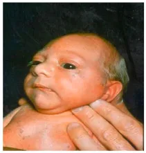
Microcephaly25
Why Check: Provides initial percentiles according to gestational age, assessing intrauterine growth. With respect to birthweight, these are categorized as: Macrosomia (>4500g); Average term neonate (2500-4500g); Low Birth Weight (1500-2500g); Very Low Birth Weight (1000-1500g); and Extremely Low Birth Weight (<1000g) – note that Very Low and Extremely Low Birth Weight infants represent about 1% of all births, but account for about 50% of all neonatal deaths. Small for gestational age (SGA) is defined as birth weight below 10th percentile for age, and may be separated out into symmetric and asymmetric as follows:
- Symmetric (head, body and length proportionally affected – 20-30% of cases): Usually cause occurs early in pregnancy, including Environmental exposures (e.g. smoking, opioids, ethanol, cocaine, phenytoin, warfarin); fetal factors (including genetic e.g. Trisomy 13, 18, 21, Turner’s Syndrome); metabolic e.g. phenylketonuria); and early infection (e.g. TORCH infections, syphilis, tuberculosis)
- Asymmetric (head circumference spared relative to ↓ length and ↓↓ weight – 70-80% of cases): Usually occurs later in pregnancy (3rd trimester), including Uteroplacental insufficiency with chronic fetal hypoxia (e.g. pre-eclampsia, hypertension, renal disease, antiphospholipid antibody syndrome; multi-gestation)
- Large for Gestational Age (LGA) is defined as birth weight above 90th percentile for age. Causes include maternal factors (e.g. maternal diabetes, maternal pre-pregnancy weight, and excess weight gain in pregnancy) and genetic syndromes.
- The Head Circumference also forms part of assessment for microcephaly or macrocephaly. Microcephaly may be familial, or associated with CNS malformations, infections (e.g. TORCH), trisomy 13 or 18, fetal alcohol syndrome or other conditions. Macrocephaly may be associated with CNS disorders, hydrocephalus or skeletal disorders, and may also be familial as an isolated phenomenon.
Pre- vs. Post-Ductal SpO2 (Congenital Heart Disease Screening)
Say: Next, I will check the baby’s oxygenation as a standard screening test for congenital heart disease.
Who to Screen: All term or late preterm newborns (born ≥34 wk GA) within 24-36 hours of age in a quiet, non-fussed state, prior to disruptive care activities (e.g. before bloodwork)
- Neonatal ICU/Special Care Nursery/Pediatric ICU babies may be screened if: cardio-respiratory status is stable and expected length of stay is <7 days, OR at time of discharge and baby is <7 days old.
Who NOT to Screen: Infants in NICU/SCN/PICU with expected length of stay >7d; Infant diagnosed with CHD prenatally (i.e. this would not be a screening exam here); Parents/guardians decline with informed consent.
Do: Measure SpO2 in RIGHT HAND and EITHER FOOT in direct sequence for 30 seconds each (once a reliable signal is obtained). Choose the highest value for each 30 second interval.
- Screen negative: SpO2 ≥95% in R Hand and Foot, and ≤3% difference between RH and F
- Screen positive: SpO2 ≤90% in RH or Foot or 3x indeterminate screens
- What to do with a positive screen: Note that +ve screen does not diagnose Congenital Heart Disease (CHD) but does warrant further workup. Initial assessment may include 4 limb blood pressures, an electrocardiogram and chest x-ray, alongside an urgent referral to pediatric cardiology followed by echocardiography to assess for Congenital Heart Disease.
- Screen indeterminate: SpO2 90-94% in RH and Foot, OR >3% difference between RH and Foot.
- Repeat screen hourly until screen negative or positive for 2 more times.
- If both 1st and 2nd rescreen are indeterminate, this is treated as a positive screen.
Report: Your baby’s pre- and post-ductal oxygen saturation screening test is normal.
Why Check26: Pre- vs. Post-Ductal SpO2 can help screen for congenital heart disease (CHD) – with incidence of 12 per 1000 live births in Canada, 25% of whom require surgery or catheter intervention in 1st year of life. While CHD can be detected prenatally, a study from in Alberta showed only 50% of newborns with CHD requiring surgery before 1 year of age were diagnosed prenatally. If CHD is identified prenatally, this would prompt referral to appropriate specialists and discussion of treatment options
Physiologically, sats in the right hand represent pre-ductal SpO2 (i.e. proximal to ductus arteriosus), while measuring sats in either foot represents post-ductal SpO2 (i.e. distal to ductus arteriosus). The ductus arteriosus typically closes within 2-3 days, hence the time-sensitive nature of screening. Note that this a screening test (i.e. asymptomatic newborns) – where patients with signs and symptoms of CHD would require a focused assessment (including those identified prenatally with CHD). Possible symptoms and signs of CHD include cyanosis, tachypnea, increased work of breathing, feeding difficulty, pounding heart, weak pulse, and lethargy. To assess for central and peripheral cyanosis, including a differential for neonatal cyanosis, please see Head and Neck above. The Pre- vs. Post-Ductal SpO2 screening test may detect cardiovascular causes of cyanosis, including27:
- CV – Cyanotic Seven (detected by pulse oximetry): Hypoplastic left heart syndrome, Pulmonary atresia with intact septum, Total anomalous pulmonary venous return, Transposition of the great arteries, Truncus arteriosus, Tetralogy of Fallot, Tricuspid atresia.
- CV – Others (usually detected by pulse oximetry, may or may not be cyanotic): Coarctation of the aorta, double outlet right ventricle, Ebstein’s anomaly, interrupted aortic arch, single ventricles, persistent pulmonary hypertension of the newborn (PPHN)
Head and Neck Exam
Head & Neck Inspection
AFMC level Pre-clerkship 1 · Clerkship 3
Say: Next, I will check the baby’s overall development and symmetry of the face.
Do: Assess the baby’s head for microcephaly, macrocephaly, and edema or hematomas. Assess the baby’s ears, nose, nostrils and mouth for dysmorphic features, including size, shape, symmetry, position, and patency. Check the eyes for signs of infection such as erythema or discharge, as well as lesions or trauma. Look for conjunctival pallor. Assess for full range of motion of the neck and look for webbed neck. Palpate for crepitus or deformity of clavicles to suggest clavicular fractures.
Report: I note no abnormalities and good symmetry of the head, ears, nose, mouth and eyes.
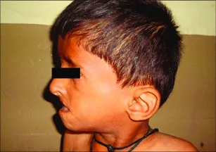
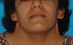
Low Set Ears27; Webbed Neck (e.g. in Turner Syndrome)28
Why Check:
- Note that moulding (change of head shape with delivery) is common and resolves in few days after birth.
- Microcephaly may be familial, or associated with CNS malformations, infections (e.g. TORCH), trisomy 13 or 18, fetal alcohol syndrome or other conditions. Macrocephaly may be associated with CNS disorders, hydrocephalus or skeletal disorders, and may also be familial as an isolated phenomenon. Edema or hematomas may represent caput succedaneum, cephalohematoma or subgaleal hemorrhage where palpation of fontanelles and sutures can help clinically differentiate them (See below).
- Facial asymmetry may indicate nerve palsy, which can be a complication of forceps use during delivery and tends to resolve spontaneously within days. Infants with facial nerve palsy may have inability to form effective seal for feeding, with drooling of feeds from paralyzed side of mouth.
- Low-set ears are associated with genetic conditions such as Down syndrome, Turner syndrome or Trisomy 18 (Edwards Syndrome), and abnormalities of the ears such as dysplastic features, sinuses, preauricular tags/pits may be associated with renal malformations.
- Eyelids may be fused before 28 weeks gestation and should open spontaneously in upright position. Erythema or discharge from the eyes may represent conjunctivitis. Conjunctival pallor may indicate fetal anemia (e.g. from acute blood loss during delivery, or chronic intrauterine process). Up-slanted palpebral fissures along with epicanthic folds are associated with Down syndrome, and abnormal distances between eyes are also associated with genetic conditions.
- Pallor may be a sign of neonatal asphyxia, shock, sepsis or anemia.
- Choanal atresia occurs when one or both nasal passages are narrowed or blocked. In bilateral choanal atresia, the infant may have cyanosis that is relieved by crying.
- Asymmetry of the mouth (asynclitism) is commonly caused by intrauterine position, resolves over time. Retrognathia (small, recessed mandible) may lead to feeding difficulty or cause respiratory obstruction.
- Webbed neck can represent Turner syndrome, and abnormal neck range of motion including head tilt can suggest neonatal torticollis (e.g. from being in fixed position in utero, or from postnatal hematoma due to birth injury). Clavicular fractures may occur in delivery process, usually asymptomatic greenstick fractures but may present with decreased movement and pain with movement of arm on affected side.
Palpation of Fontanelles and Sutures
AFMC level Pre-clerkship 1 · Clerkship 3
Say: Next, I will palpate the baby’s head for the fontanelles.
Do: Palpate with infant supine as well as upright. Feel for the anterior and posterior fontanelles, particularly the size, location and for swelling. Assess for any masses, bulges, edema or hematomas. Palpate the sutures (sagittal, coronal, lamboid, and metopic) – are they separated? If edema or hematomas are present, do they cross suture lines? Do they feel soft or firm?
Report: The anterior and posterior fontanelles and sutures appear in the appropriate position. There are no signs of swelling or bulges.
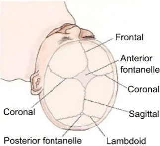
Why Check: Fontanelles should be soft and non-bulging, with anterior larger than posterior fontanelle. A full fontanelle with separated sutures is associated with increased Intracranial Pressure (↑ ICP – e.g. due to subdural hematoma, or bacterial meningitis), hypothyroidism, trisomy 13, 18 and 21, as well as bone disorders or hypophosphatemia. Small fontanelles may indicate craniosynostosis with growth restriction perpendicular to the affected sutures. For hematomas and edema, important findings include:
- Caput Succedaneum: Edema of the scalp above the periosteum over presenting part of head (e.g. prolonged engagement of fetal head in birth canal, or with vacuum-assisted delivery); crosses suture lines; resolves within few days
- Cephalohematoma: Collection of blood in subperiosteal space; does not cross suture lines; feels fluctuant, rarely expands after delivery, generally not causing significant blood loss. Generally resolves over short weeks, but calcification of hematoma and subsequent bony swelling may persist x months.
- Subgaleal Hemorrhage – Can’t Miss Diagnosis: Collection of blood between aponeurosis covering scalp and periosteum; extends across suture lines; feels firm and fluctuant; may continue to expand hours-to-days after delivery. Potential for massive blood loss, hypovolemic shock and mortality.
Red Reflex
(See Ch 40 – Ophthalmology – IF)
AFMC level Pre-clerkship 1 · Clerkship 3
Say: I will check the baby’s eyes for the red reflex and corneal light reflex.
When: Done in first 24 hours of life.
Do: To assess the Red Reflex, hold the direct ophthalmoscope to your eye and stand at a distance of 18 inches away from the child in a darkened room. Look through the ophthalmoscope to assess the red reflex. Both eyes examined simultaneously.
Report: The red reflex appears normal.
Why Check: Looking for Diminished Reflex (Congenital Cataracts, Retinoblastoma – urgent referral required), Asymmetric Reflex (strabismus, unequal/high refractive error)29. Note: Retinoblastoma and congenital cataract also cause leukocoria (white pupil in AMBIENT LIGHT or in darkened room).
Central Cyanosis
(See Ch 5 – Resp Exam – MF1)
AFMC level Pre-clerkship 4 · Clerkship 4
Say: I will check the baby for signs of good oxygenation.
Do: Gently open baby’s mouth (may be combined with checking the palate and suck reflex, see below) and inspect the tongue and mucous membranes for signs of central cyanosis.
Report: I note the baby is a healthy pink colour with no concerns for peripheral or central cyanosis.
Why Check: Cyanosis or a decrease in oxyhemoglobin saturation indicates need for rapid assessment and potential cardiopulmonary support (per PALS). A normal infant appears pink. Central cyanosis (best seen on tongue and mouth mucous membranes) suggests hypoxemia and neonatal cyanosis and is a can’t miss clinical finding. There is a long list of DDx for neonatal cyanosis, including:
- CV – Cyanotic Seven (detected by pulse oximetry)30: Hypoplastic left heart syndrome, Pulmonary atresia with intact septum, Total anomalous pulmonary venous return, Transposition of the great arteries, Truncus arteriosus, Tetralogy of Fallot, Tricuspid atresia.
- CV – Others (usually detected by pulse oximetry, may or may not be cyanotic)30: Coarctation of the aorta, double outlet right ventricle, Ebstein’s anomaly, interrupted aortic arch, single ventricles, persistent pulmonary hypertension of the newborn (PPHN - persistence of ↑ pulmonary vascular resistance, resulting in right-to-left shunting of deoxygenated blood through PFO and Ductus Arteriosus)
- Non-Cardiac Causes of Cyanosis in the Newborn (Not detected by Pre-Post Ductal SpO2 Screen):
- Pulmonary (re. Ventilation/perfusion mismatch): respiratory distress syndrome (RDS – re. surfactant deficiency), transient tachypnea of the newborn (TTN – re. retained fetal lung fluid), aspiration (of blood, amniotic fluid or meconium), pneumonia; or pneumothorax; congenital diaphragmatic hernia
- Head and Neck: Micrognathia or Retrognathia (posterior tongue causing retropharyngeal airway obstruction while infant is supine); Congenital laryngomalacia; Choanal atresia (bilateral choanal atresia may present with cyanosis that is relieved by crying)
- Neurologic (re. alveolar hypoventilation): Hypoxic ischemic encephalopathy; Intracranial hemorrhage; seizures; phrenic nerve injury; meningitis; encephalitis
- Decreased Peripheral Circulation (peripheral cyanosis): Sepsis; shock; severe hypoglycemia; hypothermia; polycythemia
- Decreased Hemoglobin O2 affinity: Methemoglobinemia
Palate Exam & Suck Reflex
Suck reflex - [1, 2]
Say: Next, I will check the baby for a normal palate and the suck reflex.
Setup: Wear a new set of sterile gloves to perform this exam (as you will be checking the suck reflex).
Do: Look inside the baby’s mouth, checking the mobility of the tongue and looking for cleft lip or palate. Look for Epstein Pearls (small white benign inclusion cysts on hard palate). Inspect for erupted teeth, which occur more frequently with cleft palate. Look for thrush (white velvety plaques with or without erythematous base on tongue and in mouth). Using your supinated 5th finger, place it in the mouth of the baby. Feel for smoothness of the palate and assess for any irregularities or cleft palate. Also, assess the baby’s reflex to suck on the finger.
Report: I note no cleft palate and the presence of the suck reflex.
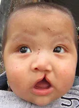
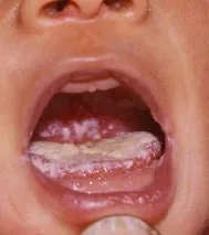
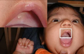
Cleft Lip/Palate31; Oral Candidiasis32; Epstein Pearls33
Why Check: Impaired tongue mobility can suggest abnormalities with frenulum and subsequent ankyloglossia (“tongue-tie”), which can lead to difficulties with feeding in the future. Cleft palate is one of the most common congenital abnormalities and warrants plastic surgery referral for repair. It is also found in newborns with trisomy 13. If untreated, cleft lip and palate can cause difficulties with feeding. Neonatal teeth are rarely seen but occur most often in lower incisor area. Epstein pearls are benign and usually disappear in a few weeks. Thrush may be asymptomatic, but some infants may have difficulties with feeding due to discomfort from lesion contact with feeding. Midline cleft lip or cleft palate may warrant further investigation of other midline defects, including the brain and thyroid. The suck reflex is a developmental (primitive) reflex, where asymmetric developmental reflexes may suggest hemiplegia or monoplegia. A functional suck reflex is important in feeding and breast latch.
Skin Exam
Peripheral Cyanosis
(See Ch 5 – Resp Exam – MF1)
AFMC level Pre-clerkship 4 · Clerkship 4
Say: Next, I will check the baby for signs of good circulation.
Do: Inspect the baby’s hands and feet for acrocyanosis as a sign of peripheral cyanosis.
Report: I note the baby is a healthy pink colour with no concerns for peripheral cyanosis.
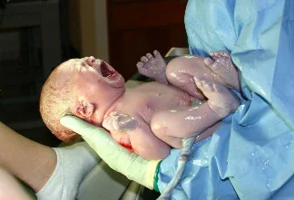
Peripheral Cyanosis34
Why Check: A normal infant appears pink. Acrocyanosis (bluish appearance of hands, feet and perioral area) is a sign of peripheral cyanosis that is common in first 48-72 hours after delivery. Note that acrocyanosis can occur as part of vasomotor instability along with cutis marmorata (pinkish blue mottling of skin when exposed to cold), telangiectasias (dilated small linear superficial blood vessels) and phlebectasia (intermittent mottling with venous prominence) are normal in premature infants. Central cyanosis (best seen on tongue and mouth mucous membranes) suggests hypoxemia and neonatal cyanosis and is a can’t miss clinical finding indicating need for rapid assessment and potential cardiopulmonary support. There is a long list of DDx for neonatal cyanosis, as detailed above (see Central Cyanosis).
Skin & Jaundice
General – [1, 3] Jaundice - [4, 4]
Say: Next, I will look at the baby’s subcutaneous fat and skin for any abnormalities or yellowing of skin.
Do: Note the amount of subcutaneous fat on anterior thighs and gluteal region as a sign of fetal nutrition. Check for abnormalities in the baby’s skin, such as rashes or skin discolorations such as pigmentation. Assess jaundice by looking for yellowing of the skin or eyes.
Report: I note no signs of cyanosis, jaundice or other abnormalities of the baby’s skin.

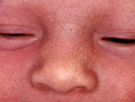

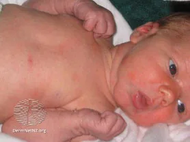
Neonatal Jaundice35; Milia36; Lanugo37; Erythema Toxicum Neonatorum38

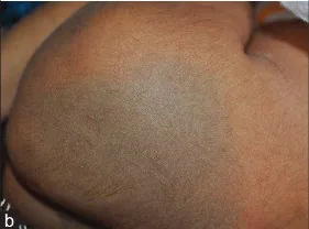
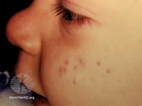
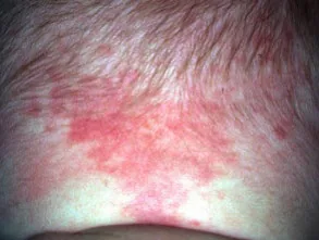
Transient Pustular Melanosis39; Congenital Dermal Melanocytosis40; Neonatal Cephalic Pustulosis41; Nevus Simplex42
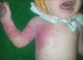
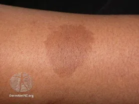
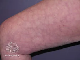
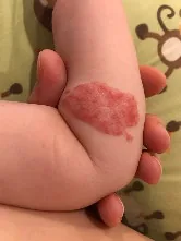
Nevus Flammeus40; Café Au Lait Macules43; Cutis Marmorata44; Infantile Hemangioma45
Why Check:
- Degree of fetal subcutaneous fat on the anterior thighs and gluteal region indicate fetal nutritional state.
- Skin Signs indicating need for rapid assessment and potential cardiopulmonary support (per PALS): Burns involving >10% of body surface area; Fever with petechiae
- Jaundice: 60% of term newborns develop jaundice, where 2% develop severe hyperbilirubinemia (>340 µmol/L in first 28 days of life). Total bilirubin (serum or capillary) is measured in the first 72 hours of life (e.g. if presenting with clinical jaundice, at time of metabolic screening test, at discharge, or by 72 hours if no other indication previously). Jaundice in first 24 hours of life is always abnormal (usually caused by hemolysis) and requires urgent evaluation. Persistent jaundice after two weeks warrants work-up. Very high bilirubin levels are associated with acute bilirubin encephalopathy and kernicterus (neuronal necrosis of basal ganglia and brainstem nuclei). Treatment with phototherapy (“Under the UV lights”) and monitoring is based on a nomogram of Postnatal age and Bilirubin (guideline)46. Causes of hyperbilirubinemia include:
- Unconjugated Hyperbilirubinemia: Physiologic neonatal jaundice (common in first 2 wks, often benign); Breastfeeding or breastmilk jaundice; ↑ bilirubin production (hemolysis, sequestered blood e.g. due to subdural hematoma, cephalohematoma, subgaleal hemorrhage, hemangioma), polycythemia, sepsis.
- Conjugated Hyperbilirubinemia: Hepatobiliary (e.g. TORCH infection, metabolic disease); Biliary atresia
- Common cutaneous findings seen in newborns to look for include:
- Lanugo (Benign): Thin hair covering skin of preterm infants, minimally present in term infants.
- Milia (Benign): White papules from retention of keratin and sebaceous material in pilaceous follicles, commonly on nose and cheeks, resolving in first few weeks of life.
- Erythema Toxicum Neonatorum (Benign): White papules 1-2mm on erythematous base; usually develop Day 2-3, more so on body than the face, and never on palms or soles. Resolves in first week.
- Transient Pustular Melanosis (Benign): Generalized superficial pustule eruption over hyperpigmented macules; pustules usually unroofed and removed in first bath so only macules remain.
- Congenital dermal melanocytosis (“Mongolian spots” - Benign): Blue-grey, greenish-blue, or brown pigmented macules with indefinite borders, due to delayed dermal melanocyte disappearance. Lesions commonly on buttocks or base of spine and may be up to 10cm in size. Usually fade by 4 years of age.
- Neonatal cephalic pustulosis (Neonatal acne): Pustular eruption on head and neck around first 3 weeks of life. Resolves spontaneously without scaring in a few months.
- Nevus simplex (“Salmon Patch, Stork bite”): Pink-red vascular capillary malformation on upper eyelid, upper lip, mid-forehead or nape of neck.
- Nevus flammeus (“Port wine stain”): Low-flow vascular capillary malformation can occur anywhere on body – may nor may not be benign (for example, occurs in Sturge-Weber disease)
- Café Au Lait Macules: Flat, uniformly hyperpigmented macules (6 or more is highly suggestive of Neurofibromatosis Type 1, and may also be a sign of McCune-Albright syndrome)
- Cutis marmorata: Reticular mottling of the skin caused by vascular response to cold, typically resolves within 1 month.
- Infantile Hemangiomas: Benign vascular tumor caused by growth of endothelial cells on blood vessels (concerning sign if found overlapping midline of spine, and if associated with a tuft of hair or large dimples above gluteal crease and anal verge re. occult spinal lesions).
Cardiovascular and Respiratory Exam
Chest Inspection
(See Ch 5 – Resp Exam – MF1)
AFMC level Pre-clerkship 4 · Clerkship 4
Say: I will now check the shape of the baby’s chest.
Do: Inspect the shape of the chest – is it small or bell-shaped? look for Pectus excavatum (abnormal concave chest shape) and Pectus carinatum (abnormal convex chest shape). Look at the baby’s nipples – are they widely spaced? Is there a supernumerary nipple (accessory nipple most often found just inferior to normal breast area)?
Report: I note the baby has a normal chest wall shape, with no abnormalities of nipple and breast tissue.
Why Check: A small thorax may occur due to pulmonary hypoplasia, and a bell-shaped thorax may indicate neuromuscular disorders. Pectus excavatum or carinatum may occur as isolated findings, or as part of clinical syndromes. Widely spaced nipples may indicate Turner Syndrome. Supernumerary nipples are usually asymptomatic but may be associated with renal anomalies.
Work and Rhythm of Breathing
(See Ch 5 – Resp Exam – MF1)
AFMC level Pre-clerkship 4 · Clerkship 4
Say: I will now assess the baby’s work and rhythm of breathing.
Do:
- Look for increased work of breathing: rapid breathing, nasal flaring, accessory muscle use (e.g. subcostal, intercostal, supraclavicular or suprasternal retractions), grunting, or gasping?
- Look for rate and rhythm of inspiratory and expiratory phases. Look at the depth of breathing. Look for signs of laboured breathing or apneas (absent breathing) during each cycle. Look for abnormal breathing patterns such as Cheyne-Stokes Breathing (periodic breathing with deep breathing gradually becoming shallower with period of apnea after – as in ↑ ICP), Shallow breathing (potential sign of pleuritic chest pain) or Kussmaul Breathing (rapid, deep laboured breathing – as in metabolic acidosis)
Report: I note no signs of increased work of breathing, with a normal rhythm of breathing and no gasping.
Why Check: Be mindful that infant breathing rate and pattern may fluctuate with sleep vs. waking, and when infant is active or crying. Note that the ribs and sternum are incompletely ossified in the newborn, causing a highly compliant chest wall – the abdomen may move outward normally during inspiration here, while the rib cage moves inward.
Increased work of breathing, irregular respirations or respiratory rate > 60 breaths/minute indicates need for rapid assessment and potential cardiopulmonary support (per PALS). Respiratory distress in the newborn is typically caused by abnormal respiratory function in transitioning from fetal to neonatal physiology. Gasping is a sign of severely impaired gas exchange requiring intervention.
In the presence of cyanosis, respiratory disease is more likely in newborns who are tachypneic and using accessory muscles of respiration. In contrast, patients with cyanosis due to congenital heart disease tend to present with mild tachypnea or hyperpnea, with less signs of increased work of breathing. Pulmonary causes (re. ventilation/perfusion mismatch) of neonatal cyanosis with signs of respiratory distress include Respiratory distress syndrome (RDS – re surfactant deficiency), transient tachypnea of the newborn (TTN – re. retained fetal lung fluid), aspiration (of blood, amniotic fluid or meconium), pneumonia; or pneumothorax; congenital diaphragmatic hernia. Another important cause of neonatal respiratory distress is persistent pulmonary hypertension of the newborn (PPHN – persistence of elevated pulmonary vascular resistance, resulting in right-to-left shunting of deoxygenated blood through PFO and Ductus Arteriosus – may also be detected by Pre- vs. Post-SpO2 screen)
Chest Palpation
(See Ch 6 – Cardiac Exam – MF1)
Heaves/Thrills - [1, 2] PMI Location - [3, 4]
Say: I will now palpate the baby’s chest to check their heart and look for any abnormalities.
Do: Palpate for the Point of Maximal Impulse using 2-3 fingers, particularly to assess for location. Using your whole hand, palpate for heaves and thrills. Palpate the chest wall for signs of crepitus.
Report: The Point of Maximal impulse is found at the Left Lower Sternal Border which is normal, with no signs of cardiac heaves or thrills, or crepitus.
Why Check: In the adult exam, the Point of Maximal Impulse (PMI) is normally found at the 5th intercostal space at the left mid-clavicular line. In newborns, the PMI is found at the Left Lower Sternal Border due to the newbown heart being right ventricle dominant (left ventricle enlarges over time – this is also reflected in how a newborn EKG may have a physiologic right axis deviation). A PMI shifted to the right may indicate Dextrocardia, or left tension pneumothorax. A PMI shifted to the left may indicate right-sided tension pneumothorax. Crepitus on palpation of the chest wall may indicate subcutaneous emphysema which can occur in pneumothorax or pneumomediastinum.
Respiratory Auscultation
(See Ch 5 – Resp Exam – MF1)
Gen. - [3, 4] Stridor (Peds) - [1, 3] Wheeze (Infant) - [1, 2]
Say: I will now listen to the baby’s lungs.
Do: Wait to auscultate until the infant is as quiet as possible. Auscultate breath sounds in bilateral lung fields. Assess for decreased or unilateral breath sounds. Listen for grunting, wheezing or stridor.
Report: The baby’s breath sounds are normal bilaterally and symmetrical.
Why Check: The Newborn exam is opportunistic – if the infant is asleep when you begin the exam, you may want to begin with auscultating the heart and lungs which is best heard when the infant is quiet. Some infants may have scattered rales for a few hours after delivery. Decreased breath sounds may represent pleural effusion, while increased breath sounds may represent consolidation or pneumonia. Asymmetrical breath sounds may represent pneumothorax. Stridor in newborns may indicate congenital disorders such as laryngomalacia, subglottic stenosis and tracheomalacia. Wheeze in infants may indicate bronchopulmonary dysplasia (presents with tachypnea, may have ↑ work of breathing), asthma/reactive airway disease, infectious (bronchiolitis, bronchitis, pneumonia, upper respiratory infection), cystic fibrosis, laryngotracheobronchomalacia, tracheoesophageal fistula, or foreign body aspiration. A wheeze in a newborn infant is more likely to be a congenital than non-congenital cause.
Cardiac Precordial Auscultation
(See Ch 6 – Cardiac and Heme Exam – MF1)
S3, S4- [1, 3] Murmur [1, 3] Abnormal S1, S2 [1, 1] Position [1, 4]
Say: Next I will listen to the baby’s heart.
Do: Auscultate the baby’s heart sounds with the bell of the stethoscope at the Aortic, Pulmonic, Tricuspid and Mitral Valves. Pay attention for abnormal heart sounds or murmurs.
Report: I note a normal S1 and normal splitting of S2. There are extra heart sounds or murmurs present.
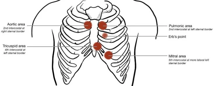
Why Check: The Newborn exam is opportunistic – if the infant is asleep when you begin the exam, you may want to begin with auscultating the heart and lungs which is best heard when the infant is quiet.
- Prominent heart sounds in the right chest may indicate dextrocardia.
- Most newborns in the first few days have murmurs that are transient and benign due to the transition from fetal to neonatal circulation. An example is the machinery-like, harsh, continuous murmur of a Patent Ductus Arteriosus (PDA) that is loudest under left clavicle and may radiate down left sternal border – tends to close after 2-3 days (re. why the Pre- and Post-Ductal O2 Sats screening test for congenital heart disease is performed within this timeframe).
- Pathologic features of murmurs prompting closer assessment: Any diastolic murmur; systolic grade ≥3; holosystolic; associated systolic click; maximal intensity at left upper sternal border; S3 or S4 gallop; worse with upright position; harsh/blowing quality; or with a friction rub.
- Innocent murmur features: Grade ≤2, Softer intensity when sitting vs. supine; short systolic duration; minimal radiation; musical or vibratory quality.
- In Newborns, relatively benign lesions tend to have loud murmurs, versus severe congenital heart disease may have no murmurs.
- In a healthy newborn, a murmur should be followed-up in a few weeks and investigated if it persists. In a critically ill newborn or with pathologic features, a murmur should be investigated promptly.
Abdomen Exam
Abdominal Inspection
(See Ch 13 – Abdo Exam – MF3)
AFMC level Pre-clerkship 3 · Clerkship 4
Say: I will inspect the abdomen for abnormalities.
Do: Observe the shape of the abdomen – does the abdomen look flat, rounded, scaphoid or protuberant? Check for any masses or bulges, hernias, distension, gastroschisis or omphalocele. Assess the umbilical cord for normal vessels (3-vessel cord with 2 umbilical arteries, vs. single umbilical artery in a 2-vessel cord), signs of infection or bleeding.
Report: There are no noticeable masses, bulges, or abnormalities of the baby’s abdomen. The umbilical cord does not appear infected or bleeding.
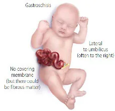
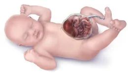
Gastroschisis47; Omphalocele48
Why Check: In newborns, a normal abdomen is slightly protuberant. A scaphoid abdomen may indicate congenital diaphragmatic hernia, while a distended abdomen may suggest intestinal obstruction, organomegaly or ascites. Note that intestinal obstruction also usually presents with bilious vomiting (re. obstruction distal to ligament of Treitz). A differential diagnosis of newborn intestinal obstruction includes:
- Intestinal atresia (common – duodenal atresia due to issue of embryogenesis, vs. jejunoileal atresia due to vascular accident in utero)
- Necrotizing Enterocolitis (life-threatening ischemic necrosis of intestinal mucosa, common in preterm)
- intestinal malrotation with volvulus (issue of embryogenesis, with intestine twisting on its mesentery – risk of bowel obstruction and infarction)
- Annular Pancreas (issue of embryogenesis, pancreas forms a circumferential mass around 2nd part of duodenum with risk of obstruction)
- Hirschsprung disease (failure of migration of inhibitory neurons to innervate distal colon, presenting with failure to pass meconium)
- Meconium ileus (failure to pass meconium – common cystic fibrosis presentation)
Umbilical hernias are common, and most resolve benignly in a few years. Gastroschisis (Bowel not covered by amnion, defect 2-3 cm lateral to umbilicus) and omphalocele (Bowel contained in sac, found in midline) require emergent surgical consultation. A single umbilical artery is associated with other congenital abnormalities, including renal conditions, prematurity and IUGR.
Abdominal Auscultation
(See Ch 13 – Abdo Exam – MF3)
AFMC level Pre-clerkship 3 · Clerkship 4
Say: I will listen to the baby’s belly.
Do: Auscultate for bowel sounds in the four quadrants, as well as overlying any bulges.
Report: Healthy bowel sounds were heard in the [quadrant].
Why Check: Abnormal bowel sounds may suggest intestinal obstruction – differential diagnosis described above. Bowel sounds heard in the chest may indicate congenital diaphragmatic hernia, and bowel sounds heard over a bulge or scrotum may suggest a hernia.
Abdominal Palpation
(See Ch 13 – Abdo Exam – MF3)
AFMC level Pre-clerkship 3 · Clerkship 4
Say: I will palpate the baby’s abdomen for any masses or enlarged organs.
Do: Palpate for abdomen for any masses, bulges, or hernias. Determine whether any hernias are reducible. Assess for organomegaly, particularly nephromegaly, hepatomegaly and splenomegaly.
What is normal: Normal liver edge 1-3 cm below R costal margin, with soft and smooth edge. The tip of the spleen may be palpable in some infants. The lower poles of both kidneys may be palpable.
Feel: There baby’s belly is soft, with no masses or bulges and no organomegaly.
Why Check: The Newborn exam is opportunistic – palpate when infant is calm, and abdomen relaxed. May flex baby’s legs or have baby suck on pacifier to promote abdominal relaxation. Umbilical hernias are common, and most resolve in a few years. It is important to assess whether hernias are reducible (↑ risk of incarceration/strangulation). Gastroschisis and omphalocele require emergent surgical consultation. An abdominal mass with projectile non-bilious vomiting may represent hypertrophic pyloric stenosis (classically weeks 3-6 of life). Masses can represent complicated meconium ileus, cysts, adrenal hemorrhage, tumours, ovarian masses, or choledochal cysts. Nephromegaly may suggest renal abnormalities (e.g. hydronephrosis, multi-cystic kidneys, renal vein thrombosis or tumours. A differential for hepatomegaly includes:
| Hyperbilirubinemia? | Splenomegaly? | Differential Diagnosis |
|---|---|---|
| Unconjugated Hyperbilirubinemia | N/A | Hemolytic anemia; Congestive heart failure; drugs; toxins |
| Conjugated Hyperbilirubinemia | Yes | TORCH infections; Bacterial infections; metabolic disorders |
| Conjugated Hyperbilirubinemia | No | Choledochal cysts; Biliary atresia (fibro-obliterative disease of extrahepatic biliary tree); neonatal hepatitis; prolonged parenteral nutrition; drugs; toxins |
| No Hyperbilirubinemia | Yes | Vascular malformation/obstruct.; liver tumor; metabolic disorder |
| No Hyperbilirubinemia | No | Tumor; malnutrition; maternal diabetes |
Genital, Hip and Femoral Pulse Exam
- Note: The infant may cry during these exams and may be best done at the end of your assessment.
Genital Exam
(See Ch 19 – Sensitive Pelvic Exams – MF3)
AFMC level Pre-clerkship 2 · Clerkship 3
Say: I will assess the development of the baby’s external genitalia, which we check for every newborn.
Do:
- For female newborns: Assess the development of labia majora, labia minora and clitoris. Check for the patency of the hymen by gentle separation of the labia minora. Assess for normal white discharge or blood, which may occur due to maternal estrogen withdrawal. Vaginal opening should be fully visible.
- For male newborns: Assess for descended testes in the scrotal sac (should be equal in size). Check for inguinal hernia or hydrocele (on transillumination of scrotum, hydrocele soft red). Retract the foreskin and assess for hypospadias (proximally displaced urethral meatus) or chordee (ventral curvature of penis). If hypospadias is present, note the location of the urethral opening.
- For all patients: Check for signs of ambiguous genitalia, including clitoromegaly, micropenis (Normal: 3.5 ± 0.7cm length, 1.1 ± 0.2cm diameter), fused labia, bilateral descended testes or bifid scrotum.
Report: I note [female] external genitalia. The labia majora, labia minora and the clitoris are appropriately developed. The hymen is patent with normal white discharge / I note [male] external genitalia. The testes have descended into the scrotal sac, and I note no signs of inguinal hernia or hydrocele. The urethra is at the distal penile shaft and gland, indicating there is no hypospadias. There are no signs of ambiguous genitalia.
Why Check:
- In female newborns, white discharge may be a normal response to estrogen withdrawal. The labia majora becomes larger as the infant approaches term. Clitoromegaly may be a sign of masculinization, virilizing tumor, maternal androgen production or maternal drug use.
- In male newborns, undescended testes (unilateral or bilateral) may occur, more common in preterm patients. Testes descend before 6 months of age in most cases –persistent cryptorchidism ↑ risk of malignancy. Inguinal hernias may represent patent processus vaginalis, and often require surgical repair re. risk of incarceration and strangulation. Hydrocele occurs when the patent processus vaginalis allows only fluid to pass through, typically resolves spontaneously. Patients with hypospadias require a urology consultation and should not undergo circumcision as the foreskin may be used for repair.
- Ambiguous genitalia may suggest chromosomal abnormalities. In XY patients, this can suggest Androgen Insensitivity Syndrome, 5a reductase deficiency, etc. In XX patients, this can suggest congenital adrenal hyperplasia, ovotesticular disorder, aromatase deficiency, etc. These chromosomal abnormalities may influence sex presentation and affect fertility in later life. This is a medical emergency as potential signs of adrenal or pituitary dysfunction (e.g. congenital adrenal hyperplasia).
Sacrum and Anus
AFMC level Pre-clerkship 1 · Clerkship 3
Say: I will assess the patency of the anus and for any sacral abnormalities, which we check for every newborn.
Do: Assess for anal patency (particularly if infant has not passed meconium). Check for a sacral dimple or tuft of hair (hypertrichosis) about an inch from the anal verge.
Report: I note that the anus appears patent, and there is no dimple or tuft of hair in the sacrum.
Why Check: An imperforate anus is associated with trisomy 18 and 21, as well as VACTERL syndrome (vertebral defects, anal atresia, cardiac defects, tracheo-esophageal fistula, renal anomalies, and limb abnormalities). A sacral dimple or hair tuft may suggest spinal dysraphism or spina bifida – particularly concerning if large (>0.5cm in size), above the gluteal cleft (>2.5cm from anal verge), with tuft of hair (hypertrichosis) or discolouration.
Hip Exam (Barlow & Ortolani Maneuvers)
(See Ch 27 – Hip Exam – MF4)
AFMC level Pre-clerkship 1 · Clerkship 3
Say: I will check the baby’s hip joints for stability, which we check for every newborn.
Do: Infant supine on examination table with diaper off.
- Barlow Maneuver: Stabilize infant’s pelvis with one hand and grasp abducted and flexed thigh with other hand. Hip flexed to 90 degrees. Adduct the hip with an additional posterior force on the anterior hip. A positive test is felt as a clunk.
- Ortolani Maneuver: If a clunk is felt on Barlow Maneuver, Abduct the hip with an additional anterior force on the posterior thigh. A positive test is the reduction of the dislocation (may also be felt as a clunk). Repeat with contralateral hip.
Report: The hip joints are stable on each side – Barlow and Ortolani screen is negative for Developmental Dysplasia of the Hip, which is reassuring.
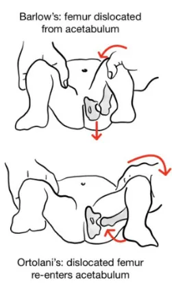
Why Check: For infants delivered by breech presentation, the Ortolani and Barlow Maneuvers have a 66.7% sensitivity and 95.8% specificity for detecting developmental dysplasia of the hip (DDH). DDH requires referral to a pediatric orthopedic surgeon and correction. The sensitivity of test is lowered in an agitated, crying infant. Risk factors for DDH include ligamentous laxity, female, positive FMHx, breech presentation, primiparous parent, oligohydramnios & postnatal infant position.
Central Pulses (Femoral)
AFMC level Pre-clerkship 1 · Clerkship 2
Say: I will palpate the baby’s pulse in the diaper area to check circulation, which we check for every newborn.
Do: Using your first two fingers, assess for the femoral pulse, located midway between ASIS and symphysis pubis. Particularly note the strength of the femoral pulse, as well as the rhythm and regularity.
Report: Femoral pulse is 2+, with regular rate and rhythm.
Why Check: Poor perfusion with weak or absent distal pulses indicates need for rapid assessment and potential cardiopulmonary support (per PALS). This exam is best completed along with the genital and hip exam within a regional approach. Note the infant may cry during these exams and may be best done at the end of your assessment. A diminished or absent femoral pulse may represent ductal-dependent cardiac lesions. This can include coarctation of the aorta, which may be seen with cyanosis of the legs but not of the arms. Weak pulses can represent poor cardiac output, such as aortic stenosis, while bounding pulses can represent high cardiac output, such as patent ductus arteriosus. If the femoral pulses are abnormal, a more complete pulse exam should be performed, including the brachial, radial, and pedal pulses. For details about palpation for central and peripheral pulses, see Ch 8 – Peripheral Vascular Exam – MF1.
Musculoskeletal and Neurologic Exam
- Note: Neurologic signs indicating need for rapid assessment and potential cardiopulmonary support (per PALS) include:
- Altered level of consciousness (unusual irritability or lethargy, or failure to respond to parents or painful procedures)
- Seizures
Extremities
AFMC level Pre-clerkship 1 · Clerkship 3
Say: I will assess the baby’s arms and legs for any abnormalities.
Do: Assess the extremities for development, symmetry, full range of movement. See if the baby is spontaneously moving the extremities (indicating muscle tone) Check for syndactyly (fusion of digits) or polydactyly (extra digits). Check the hands for a single palmar crease or clinodactyly (incurving of 5th finger). Assess for brachial plexus palsies, such as Erb palsy (e.g. C5-C7 palsy “Waiter’s Tip sign” – adduction and internal rotation of arm, extension and pronation of forearm, and flexion of wrist and fingers) or Klumpke palsy (“claw hand” isolated hand/wrist paralysis and absent grasp reflex and abnormal Moro Reflex re. hand not opening). Assess the feet for talipes equinovarus (clubfoot).
Report: I note no extra or missing digits. There is no signs of a palsy or clubfoot. The baby is moving its extremities spontaneously, indicating good muscle tone, as well as full range of movement.
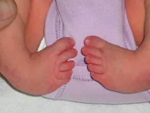
Talipes equinovarus (Clubfoot)49
Why Check: Polydactyly is typically benign, but often removed for cosmetic reasons. Single palmar creases are associated with trisomy 21 but are non-specific. Clinodactyly may suggest Trisomy 21 or Turner syndrome. Clubfoot is a common orthopedic abnormality and requires referral to an orthopedic surgeon. Erb’s Palsy is the most common brachial plexus injury and indicates brachial plexus injury to the C5-C7 roots. Alternatively, Klumpke’s Palsy is rarer and indicates brachial plexus injury to the C8-T1 roots with isolated hand paralysis and associated ipsilateral Horner Syndrome (explored in Ch 31 – Screening Neuro Exam – MF4). Clavicular fractures may present with ↓ movement of the arm on the affected side.
Passive Tone and Posture
AFMC level Pre-clerkship 1 · Clerkship 2
Say: I will assess the baby’s resting muscle tone.
Do: Assess the baby’s resting neonatal posture with head placed midline. Resting posture should be symmetric in all 4 extremities and can be measured with resistance to passive movement of the limbs. The normal resting posture varies with gestational age, as follows:
| Gestational Age | Normal Findings |
|---|---|
| ≤28wks | All 4 extremities passively extended |
| 32wks | Slight flexion at knee, then hip; upper extremities extended |
| 34wks | Increasing knee and hip flexion; upper extremities extended |
| 36wks | Weak flexion of all 4 extremities |
| 40wks | Strong flexion of all 4 extremities |
Report: There is normal resting muscle tone, with no spasticity or rigidity indicative of a palsy.
Why Check:
- Hypotonia in the newborn can be due to systemic illness causing the infant to appear ill (e.g. sepsis, TORCH infection, congenital heart disease). Otherwise, it is a feature of a number of genetic syndromes (e.g. Trisomy 13, 18, 21; and Prader-Willi Syndrome); inborn errors of metabolism; endocrine disorders (e.g. congenital adrenal hyperplasia, congenital hypothyroidism, hypoglycemia); early cerebral palsy (progresses to spasticity/dystonia) or primary neuromuscular disorders (e.g. hypoxic-ischemic myelopathy, myopathy, neuromuscular junction, peripheral neuropathy, anterior horn cell disorders).
- Hypertonia in the newborn is due to spasticity or rigidity. It is typically due to hypoxic-ischemic encephalopathy, cerebral palsy, or congenital brain lesions.
Developmental (Primitive) Reflexes50
AFMC level Pre-clerkship 1 · Clerkship 2
Say: I will assess the baby and gently move the baby around to check for reflexes present in infancy.
Do: Holding infant well-supported, perform each of the developmental reflexes as detailed below:
| Reflex | Description | Age of First Appearance | Age of Expected Disappearance |
|---|---|---|---|
| Moro (Startle) | Sudden dropping of infant’s head in relation to trunk; expect abduction & extension of infant arms, opening hands, then flexion in all 4 limbs | 34-36 Weeks PCA | 6 months |
| Stepping | Hold infant in vertical position with feet in contact on a flat surface; expect slow alternating stepping flexion & extension of legs | 32 Weeks PCA | 2 months |
| Palmar | Place a finger in infant’s open palm. Infant will close their hand around your finger and tighten grip on attempting to withdraw your finger. | 38-40 Weeks PCA | 6 months |
| Plantar | Place a finger under infant’s toes. Infant will plantarflex toes to curl around your finger | 38-40 Weeks PCA | 15 months |
| Galant (Trunk Incurvation) | Place infant in prone position, and stroke thoracolumbar paravertebral region unilaterally; expect movement of trunk & hips toward stimulus | 38-40 Weeks PCA | 4 months |
| Asymmetric Tonic Neck Reflex (ATNR) | Turn head and neck to the left; expect extension of left upper & lower limbs, and flexion of the right upper limb (Fencing posture)> | 38-40 Weeks PCA | 3 months |
| Rooting & Sucking | Stroke infant’s cheek. Infant will turn head towards the motion and begin to make sucking motions. | 38-40 weeks’ gestation | 3 months |
Report: I note normal developmental reflexes for age, with good strength and tone also noted.
Why Check: Asymmetric developmental reflexes may suggest hemiplegia or monoplegia. Developmental reflexes are developmentally expected and disappear with maturing cortical inhibitory functioning. If lasting beyond 12 months, they raise suspicion of cerebral palsy and other etiologies. They are also, technically, frontal release signs (See Chapter 31 – Screening Neuro Exam – MF4) although many are not practical to elicit in adult patients. NOTE: PCA = Post-Conceptional Age.29
Postural Reactions50
AFMC level Pre-clerkship 1 · Clerkship 2
Say: I will gently move the baby around to check postural reactions for tone and strength.
Do: Holding infant well-supported, perform each of the postural reaction tests as detailed below:
| Postural Reactions | Description |
|---|---|
| Ventral Suspension | Hold infant well-supported in a prone position with your hands supporting their anterior chest & abdomen. Expect head erect, extension of spine, flexion at elbows & hips & knees & ankles with adequate tone to not drape limp over examiner’s hands. Measures strength of trunk & neck. |
| Vertical Suspension | Hold infant well-supported at axillae B/L without grasping thorax and lift straight up. Expect head midline, flexion at hips & knees & ankles with adequate truncal resistance to not fall through examiner fingers. Measures strength of shoulder girdle. |
| Traction Response | Infant begins in supine position. Grasp infant hands bilaterally and pull towards a sitting position. Normal traction is felt by examiner as infant pulling back against traction, flexion at elbows & knees and ankles. Infant able to keep head in line with trunk 1-2s on pulling from supine to sitting. Measures strength of trunk & neck. |
Report: I note normal postural reaction tests with good strength and tone.
Postural reactions: Postural reactions test active motor tone and strength. By 32-34 weeks gestational age, infants should exhibit symmetric, smooth and spontaneous movements in all 4 extremities. Along with passive tone/posture testing, postural reactions form a core part of the motor exam for newborns and can help identify hypotonia or hypertonia (due to spasticity or rigidity). As noted, more focal injuries such as brachial plexus injuries and clavicular fractures can affect the results of the exam.
Part 4: Connections to Specific Chapters in the Text
For the sake of a systems approach to the examination, pediatric exam maneuvers that are systems-specific are described in their corresponding chapters. The following are connections to these maneuvers in the text:
References
-
American Academy of Pediatrics, Committee on Fetus and Newborn; American College of Obstetricians and Gynecologists and Committee on Obstetric Practice. The Apgar score. Pediatrics. 2006;117(4):1444-1447. ↩ ↩2
-
Harlor AD Jr, Bower C; Committee on Practice and Ambulatory Medicine, Section on Otolaryngology-Head and Neck Surgery. Hearing assessment in infants and children: recommendations beyond neonatal screening. Pediatrics. 2009;124(4):1252-1263. ↩
-
Accessed June 21, 2022. http://breastfeedingresourcesontario.ca/sites/default/files/pdf/Res_Strategy_IDM_Toolkit_EN.pdf ↩
-
Bayoglu BU, Bakar EE, Kutlu M, Karabulut E, Anlar B. Can preschool developmental screening identify children at risk for school problems? Early Hum Dev. 2007;83(9):613-617. ↩
-
Panagiotou L, Rourke LL, Rourke JT, Wakefield JG, Winfield D. Evidence-based well-baby care. Part 1: overview of the next generation of the Rourke Baby Record. Can Fam Physician. 1998;44:558-567. ↩ ↩2
-
Rosenbaum P, et al. The ‘F-words in childhood disability: I swear this is how we should think! Child Care Health Dev. 2012;38(4):457-463. ↩
-
Canadian Medical Protective Association. Accessed August 17, 2019. https://www.cmpa-acpm.ca/en/advice-publications/browse-articles/2015/when-to-disclose-confidential-information ↩ ↩2
-
Canadian Medical Protective Association. Accessed August 17, 2019. https://www.cmpa-acpm.ca/en/advice-publications/browse-articles/2019/hit-the-brakes-do-you-need-to-report-your-patients-fitness-to-drive ↩ ↩2
-
College of Physicians and Surgeons of Ontario. Accessed August 17, 2019. https://www.cpso.on.ca/Physicians/Policies-Guidance/Policies/Mandatory-and-Permissive-Reporting#child ↩
-
Ontario Association of Children’s Aid Societies. Accessed August 17, 2019. https://www.oacas.org/childrens-aid-child-protection/duty-to-report/ ↩
-
Accessed August 17, 2019. http://www.children.gov.on.ca/htdocs/English/childrensaid/reportingabuse/abuseandneglect.aspx ↩
-
Ontario Association of Children’s Aid Societies. Accessed August 17, 2019. http://www.oacas.org/childrens-aid-child-protection/locate-a-childrens-aid-society/ ↩
-
Recommendations. Choosing Wisely Canada. Accessed August 1, 2022. https://choosingwiselycanada.org/recommendations/#list ↩
-
Shah S, Bachur R, Kim D, Neuman MI. Lack of predictive value of tachypnea in the diagnosis of pneumonia in children. Pediatr Infect Dis J. 2010;29(5):406-409. ↩
-
Ogedegbe G, Pickering T. Principles and techniques of blood pressure measurement. Cardiol Clin. 2010;28(4):571-586. ↩
-
Grummer-Strawn LM, Reinold C, Krebs NF; Centers for Disease Control and Prevention (CDC). Use of World Health Organization and CDC growth charts for children aged 0-59 months in the United States. MMWR Recomm Rep. 2010;59(RR-9):1-15. ↩
-
Accessed March 13, 2019. http://bit.ly/2w71Up6 ↩
-
Accessed October 2, 2019. http://med.stanford.edu/content/dam/sm/pediatricsclerkship/documents/5-Developmental-Milestones-MedU.pdf ↩
-
Canadian Task Force on Preventive Health Care. Recommendations on screening for developmental delay. CMAJ. 2016;188(8):579-587. ↩ ↩2 ↩3
-
Fuloria M, et al. The newborn examination: part I. Emergencies and common abnormalities involving the skin, head, neck, chest, and respiratory and cardiovascular systems. Am Fam Physician. 2002;65:61-68. ↩
-
Fuloria M, Kreiter S. The newborn examination: part II. Emergencies and common abnormalities involving the abdomen, pelvis, extremities, genitalia and spine. Am Fam Physician. 2002;65:265-270. ↩
-
Lewis ML. A comprehensive newborn examination: part I. General, head and neck, cardiopulmonary. Am Fam Physician. 2014;90(5):289-296. ↩
-
Lewis ML. A comprehensive newborn examination: part II. Skin, trunk, extremities, neurologic. Am Fam Physician. 2014;90(5):297-302. ↩
-
Weiner GM. Textbook of Neonatal Resuscitation. 7th ed. American Academy of Pediatrics; 2016. ↩ ↩2 ↩3
-
Hanzlik E, Gigant J. Microcephaly. Children. 2017;4(6):47. ↩
-
Narvey M, et al. Pulse oximetry screening in newborns to enhance detection of critical congenital heart disease. Paediatr Child Health. 2017;22(8):494-498. ↩
-
Krishnamurthy B, et al. Structural anomaly of chromosome 18 with Edward syndrome phenotype. Int J Contemp Pediatr. 2014;1(3):181-183. ↩ ↩2
-
File:Preoperative webbed neck in Turner syndrome.jpg. Wikimedia Commons. Accessed April 1, 2023. https://commons.wikimedia.org/wiki/File:Preoperative_webbed_neck_in_Turner_syndrome.jpg ↩
-
American Academy of Pediatrics, Section on Ophthalmology. Red reflex examination in neonates, infants, and children. Pediatrics. 2008;122(6):1401-1404. ↩ ↩2
-
Accessed June 21, 2022. https://www.newbornscreening.on.ca/sites/default/files/cchd_training_presentation.pdf ↩ ↩2
-
File:Cleftlipandpalate.JPG. Wikimedia Commons. Accessed May 2, 2023. https://commons.wikimedia.org/wiki/File:Cleftlipandpalate.JPG ↩
-
NHS. Accessed April 1, 2023. https://www.nhs.uk/conditions/oral-thrush-mouth-thrush/ ↩
-
File:Epstien pearl.png. Wikimedia Commons. Accessed April 1, 2023. https://commons.wikimedia.org/wiki/File:Epstien_pearl.png ↩
-
File:HumanNewborn.JPG. Wikimedia Commons. Accessed April 1, 2023. https://commons.wikimedia.org/wiki/File:HumanNewborn.JPG ↩
-
NHS. Accessed April 1, 2023. https://www.nhs.uk/conditions/jaundice-newborn/symptoms/ ↩
-
Milium. DermNet. Accessed April 1, 2023. https://dermnetnz.org/topics/milium ↩
-
File:Lanugo.png. Wikimedia Commons. Accessed April 1, 2023. https://commons.wikimedia.org/wiki/File:Lanugo.png ↩
-
Toxic erythema of the newborn. DermNet. Accessed April 1, 2023. https://dermnetnz.org/topics/toxic-erythema-of-the-newborn ↩
-
Transient neonatal pustular melanosis. DermNet. Accessed April 1, 2023. https://dermnetnz.org/topics/transient-neonatal-pustular-melanosis ↩
-
Sadana DJ, et al. A clinical and statistical survey of cutaneous changes in the first 120 hours of life. Indian J Dermatol. 2014;59(6):552-557. ↩ ↩2
-
Infantile acne. DermNet. Accessed April 1, 2023. https://dermnetnz.org/topics/infantile-acne ↩
-
Naevus simplex. DermNet. Accessed May 2, 2023. https://dermnetnz.org/topics/naevus-simplex ↩
-
Neurofibromatosis images. DermNet. Accessed April 1, 2023. https://dermnetnz.org/topics/neurofibromatosis-images ↩
-
Cutis marmorata. DermNet. Accessed April 1, 2023. https://dermnetnz.org/topics/cutis-marmorata ↩
-
File:Infantile hemangioma.jpg. Wikimedia Commons. Accessed May 2, 2023. https://commons.wikimedia.org/wiki/File:Infantile_hemangioma.jpg ↩
-
Canadian Paediatric Society. Accessed June 15, 2025. https://cps.ca/en/documents/position/hyperbilirubinemia-newborns ↩
-
Centers for Disease Control and Prevention. Accessed April 1, 2023. https://www.cdc.gov/ncbddd/birthdefects/gastroschisis.html ↩
-
Centers for Disease Control and Prevention. Accessed April 1, 2023. https://www.cdc.gov/ncbddd/birthdefects/omphalocele.html ↩
-
File:Pied bot, varus équin (bilateral).jpg. Wikimedia Commons. Accessed May 2, 2023. https://commons.wikimedia.org/wiki/File:Pied_bot,_varus_%C3%A9quin_(bilateral).jpg ↩
-
Zafeiriou DI. Primitive reflexes and postural reactions in the neurodevelopmental examination. Pediatr Neurol. 2004;(31):1-8. ↩ ↩2


