Medical Foundations 2
Chapter 9Volume Status Exam
Part 1: Background – Key Physiological Principles and Terminology
Key Physiology Principles
Fluid compartments
Total Body Water ~60% of body mass
- 2/3 Intracellular fluid (ICF)
- 1/3 extracellular fluid (ECF)
- 3/4 interstitial
- 1/4 plasma (Intravascular)
ICF and ECF are separated by capillary walls and cell membranes.
- ICF: Potassium = dominant cation
- ECF: Sodium = dominant cation
- Note: Total Body Water may be ↓ in elderly and with ↑ BMI; it may be ↑ in neonates and with ↓ BMI.
Osmotic pressure – pressure gradient due to solute concentration differences across a semipermeable membrane. Involved in ICF vs. ECF fluid exchange.
Starling Forces
Starling forces determine net flow of fluid between interstitium and plasma.
- Hydrostatic pressure – pressure of fluid on vessel walls
- Colloid (oncotic) pressure – osmotic pressure gradient generated by proteins (especially albumin)
Regarding capillary (intravascular):
- Capillary hydrostatic pressure (Pc) – pushes fluid out
- Interstitial hydrostatic pressure (Pi) – pushes fluid in
- Capillary colloid (oncotic) pressure (πc) – pulls fluid in
- Interstitial colloid (oncotic) pressure (πi) – pulls fluid out
Starling Equation: Net flow = Kf [(Pc - Pi) - σ(πc - πi)]
- Kf = coefficient for vessel permeability for fluid
- σ = coefficient for vessel permeability for protein
- Key takeaway: Capillary fluid exchange determined by hydrostatic pressure gradient (Pc - Pi) and oncotic pressure gradient (πc - πi), mediated by vessel permeability.
Filtration OUT occurs on arterial side of capillary bed (high Pc), and absorption IN occurs on venous side (πc ↑ as intravascular fluid and Pc ↓).
Edema: Usually due to ↑ capillary hydrostatic pressure (e.g. CHF, Na retention) or ↓ capillary oncotic pressure (e.g. nephrotic syndrome, cirrhosis).
Hypervolemia vs. Hypovolemia
Hypervolemia
Definitions:
- Clinical congestion: ↑left ventricular diastolic pressure (LVDP), leading to pressure backup, causing symptoms of fluid overload due to increased hydrostatic pressure in Starling equation (e.g. dyspnea, crackles, peripheral edema, JVD)
- Hemodynamic congestion: ↑ LVDP without overt fluid overload symptoms, often precedes clinical congestion by days to weeks. Indicates clinical manifestations are imminent
Causes:
- ↑ Sodium/Water Intake: e.g. IV Fluids; ↑Na+ intake
- ↑ Sodium/Water Retention: e.g. Liver cirrhosis; CHF; Nephrotic syndrome; Renal failure
Hypovolemia
Absolute hypovolemia
- Definitions: External loss of fluid from the body
- Causes: Trauma, overt bleeds, GI bleeds (upper or lower), burns, severe diarrhea/vomiting, excess urination (e.g. polyuria in diabetes)
Relative hypovolemia
- Definitions: Fluid shift from the intravascular to the extravascular space (AKA “third-spacing”). Occurs due to changes in the Starling equation (e.g. decreased oncotic pressure).
- Causes: Distributive shock (e.g. sepsis, neurogenic shock, anaphylactic shock); hypoalbuminemia, hyponatremia
Can’t Miss Diagnosis – Shock
- Definition: Shock is a state of cellular and tissue hypoxia due to ↓ oxygen delivery, ↑ oxygen consumption, or inadequate oxygen utilization. Shock is a can’t-miss clinical diagnosis. Early recognition can reduce risk of irreversible end-organ dysfunction.
- Key Idea: In managing shock, general goal is to maintain a MAP of ≥ 65 mmHg.
- MAP goal may be individualized depending on cause of shock (e.g. MAP goal higher for neurogenic shock to maintain cerebral perfusion pressure), comorbidities, etc.
- True MAP measured with direct arterial pressure wave monitoring e.g. Arterial line.
- Determinants of Mean Arterial Pressure (see diagram to right) help explain mechanisms underlying different etiologies of shock. These are explored in the table below.
- Note: Students are not expected to know in pre-clerkship the details about management of shock (e.g. inotropes and vasopressors). This is included for reference. You may explore this further in clerkship and residency (e.g. Emergency Medicine, Intensive Care Unit, Critical Care rotations)
Mean Arterial Pressure (MAP) ≈ 1/3 Systolic BP (SBP) + 2/3 Diastolic BP (DBP)
Mean Arterial Pressure (MAP) = Cardiac Output (CO) × Systemic Vascular Resistance (SVR)
Cardiac Output (CO) = Heart Rate (HR) × Stroke Volume (SV)
Types of Shock and Etiologies
Hypovolemic Shock (issue of ↓ preload)
- Fluid loss (e.g. diarrhea, diuresis, burns)
- Hemorrhage (e.g. trauma, overt bleed, GI bleed, surgical bleed, ruptured abdominal aortic aneurysm)
| HR | SVR | JVP | Extremities | Treatment |
|---|---|---|---|---|
| ↑ | ↑ | ↓ | Cold, clammy | IV Fluids, Blood products |
Cardiogenic Shock (intracardiac causes of cardiac pump failure)
- Cardiomyopathic: Myocardial ischemia, Cardiomyopathy, Congestive heart failure
- Arrhythmogenic: Includes Tachyarrhythmia (e.g. A-Fib/Flutter, AVNRT, Ventricular tachycardia, V-Fib) and Bradyarrhythmia (e.g. complete heart block, Mobitz Type II second degree heart block)
- Mechanical (Severe valvulopathy e.g. Aortic stenosis, aortic regurgitation, mitral stenosis, ventricular septal defect, hypertrophic cardiomyopathy)
| HR | SVR | JVP | Extremities | Treatment |
|---|---|---|---|---|
| Variable | ↑ | ↑ | Cold, clammy | No IV Fluids, Inotropes, Pressors, diuretics (e.g. in CHF) |
Obstructive Shock (usually extracardiac causes of cardiac pump failure)
- Pulmonary vascular: Massive pulmonary embolism, Severe pulmonary hypertension
- Mechanical: Tension pneumothorax, cardiac tamponade
| HR | SVR | JVP | Extremities | Treatment |
|---|---|---|---|---|
| ↑ | ↑ | ↑ | Cold, clammy | IV Fluids |
Distributive Shock (issue of ↓ systemic vascular resistance)
- Septic: e.g. Gram-positive vs. gram-negative bacteria; viral; fungal
- Non-septic: e.g. Neurogenic (spinal cord injury, traumatic brain injury, neuraxial anesthesia); Anaphylaxis (allergen exposure); Adrenal crisis
| HR | SVR | JVP | Extremities | Treatment |
|---|---|---|---|---|
| Neurogenic ↓ Others ↑ | ↓ | ↓ | Warm, flushed | IV Fluids, Pressors |
- To manage shock, we identify and treat reversible causes if possible and within patient goals of care, for example:
- Achieving hemostasis e.g. through surgical intervention in hemorrhagic shock
- Cardiac cath lab for percutaneous coronary intervention (PCI) for myocardial infarction causing cardiogenic shock
- Pacing a patient with bradyarrhythmia; or shocking a patient with tachyarrhythmia causing cardiogenic shock
- Consideration of Prosthetic valve replacement for severe valvulopathy causing cardiogenic shock
- Thrombolysis for patient with massive pulmonary embolism causing obstructive shock
- Needle decompression (at 2nd intercostal space, midclavicular line) or chest tube thoracostomy (at 5th intercostal space, mid-axillary line) for tension pneumothorax causing obstructive shock
- Pericardiocentesis for patient with acute cardiac tamponade causing obstructive shock
- Broad spectrum antibiotics for a patient with sepsis causing distributive shock
- Epinephrine for a patient with anaphylaxis causing distributive shock
- Neurosurgical consultation for a patient with traumatic brain injury causing distributive shock
Part 2: Focused History – Based on Chief Complaint (Begin with Open-Ended Questions)
HPI, Assoc. Symptoms
Hypervolemia
- Dyspnea: A subjective experience of uncomfortable breathing
- Focus on Exertion i.e. SOB on Exertion (SOBOE):
- Do you find you become short of breath when walking up a flight of stairs?
- How many flights of stairs? How many blocks can you walk before you need to rest?
- Do you find you are short of breath at rest? – Red Flag Symptom
- Note: Refer to New York Heart Association (NYHA) CHF classifications or MMRC Breathlessness Scale for more info.
- Focus on Position re. Orthopnea (Breathlessness sensation worse when laying down, better sitting up – occurs immediately when lying flat due to increased preload to heart): Is SOB worse when lying down, better when siting up? Are you able to lay flat? How many pillows do you sleep on?
- Focus on Paroxysmal Nocturnal Dyspnea (PND – SOB awakening patient after 1-2 hours of sleep – occurs due to ↑ volume from lymphatic resorption of edema, takes longer than orthopnea): Do you wake up in the middle of the night gasping for air or feeling like you can’t breathe? Does it get better after sitting up?
- Focus on Exertion i.e. SOB on Exertion (SOBOE):
- Edema: Swelling of tissues – most commonly in dependent areas e.g. lower extremities
- Recent Weight Gain: Intentional? Any changes in your lifestyle that could explain it?
- Diet – ↑ Sodium intake and/or Fluid intake: Patients prone to volume overload (e.g. in heart failure) are often salt and/or fluid restricted as part of the management plan
- Medication Adherence: Non-adherence to medications (e.g. diuretics) can tip patients into volume overload
- Associated symptoms for common etiologies:
- Congestive heart failure (common cause of hypervolemia): SOBOE, orthopnea, PND, peripheral edema, nocturia, fatigue, palpitations
- Renal failure: Oliguria/anuria (re. ↓ urine output), Insomnia, pruritus, fatigue, edema, nausea/vomiting, weight loss, anorexia.
- Nephrotic syndrome: Frothy urine, generalized edema (e.g. including in non-dependent areas e.g. periorbital edema), ascites, thrombotic events (e.g. stroke, pulmonary embolism), fatigue, recent infections (e.g. cellulitis)
- Liver cirrhosis (Explored in Ch 13 – Abdo Exam – MF3): Weakness, fatigue, weight loss, anorexia; jaundice, pruritus, easy bruising
- Decompensating events: Hepatic encephalopathy (presenting with confusion, sleep-wake cycle reversal, asterixis); Ascites; Esophageal varices (presenting with hematemesis, melena or hematochezia); Hepatorenal syndrome (presenting with oliguria re. ↓ urine output); Spontaneous bacterial peritonitis (presenting with fever, abdominal pain, confusion)
- Relevant Equation: Sodium deficit in hyponatremia (NOTE: hyponatremia is usually an issue of water rather than sodium)
Hypovolemia
- Symptoms of volume depletion: pre-syncope, syncope, orthostatic dizziness
- Symptoms of shock: Light-headedness, altered level of consciousness (agitation, disorientation, lethargy, stupor, coma), oliguria, cold/clammy vs warm/flushed, dry mucus membranes
- Ask about relevant causes of absolute hypovolemia: Trauma, overt bleeding, burns, upper GI bleeds (melena stools, hematemesis, coffee ground emesis), lower GI bleeds (hematochezia, bright red blood per rectum), recurrent diarrhea/vomiting (ask for recent antibiotics re: C. diff), increased urination (polyuria), iatrogenic (diuretic dose)
- Ask about relevant causes of relative hypovolemia: Recent infection (progression to sepsis, or septic shock), anaphylaxis/allergies, neurogenic shock (e.g. traumatic brain injury, spinal cord injury), liver disease (re. hypoalbuminemia causing ↓ capillary oncotic pressure)
- Recent Weight Loss: Intentional or unintentional?
- Diet – ↓ Sodium and/or fluid intake: Insufficient oral salt and/or water intake (e.g. due to altered mental status)
- Relevant Equation: Free water deficit in hypernatremia (NOTE: hypernatremia is usually an issue of water rather than sodium)
Past Medical History
Hypervolemia
- Cardiac: Diseases Heart failure (very common cause of hypervolemia), coronary artery disease (CAD, e.g. MI, angina), arrhythmias, valvular heart disease, cardiomyopathies.
- Chronic Kidney Disease (CKD): Diabetes and hypertension are two most common causes of CKD
- Polycystic Kidney Disease: Includes autosomal dominant and autosomal recessive types.
- Liver Cirrhosis: Explored further in Ch 13 – Abdo Exam – MF3
- Common Etiologies: Non-alcoholic fatty liver disease (NAFLD), alcohol-related, viral hepatitis (especially hepatitis B and hepatitis C)
- NAFLD: Spectrum of disease including fatty liver (NAFLD), non-alcoholic steatohepatitis (NASH) and cirrhosis. NAFLD is a feature of metabolic syndrome (along with obesity, insulin resistance, hypertension, hypertriglyceridemia and low HDL)
- Other Etiologies: Hemochromatosis; Autoimmune hepatitis; primary biliary cirrhosis; primary sclerosing cholangitis; Wilson disease; Alpha-1 antitrypsin deficiency; Congestive hepatopathy
- Common Etiologies: Non-alcoholic fatty liver disease (NAFLD), alcohol-related, viral hepatitis (especially hepatitis B and hepatitis C)
Relevant clinical scores:
- Light’s Criteria – determines exudative vs. transudative pleural effusion
- NYHA HF Classification – stratifies severity of heart failure by symptoms
- Modified Medical Research Council (mMRC) Dyspnea Scale – stratifies severity of dyspnea in respiratory diseases
- MELD Score – Stratifies severity of end-stage liver disease
- Child-Pugh Score for Cirrhosis Mortality – Estimates cirrhosis severity
- Kidney Failure Risk Calculator – Predicts 5-year risk of progression to kidney failure in patients with chronic kidney disease (CKD)
Hypovolemia
- GI (explored in Ch 13 – Abdo Exam – MF3):
- Liver disease: portal hypertension ↑ risk of varices (abnormally dilated, swollen blood or lymph vessel, e.g. esophageal varices) → ↑ GI bleed risk
- GI bleeds: upper or lower, etiology
- Vomiting: gastritis/esophagitis (vomiting)
- Diarrhea: gastroenteritis, IBD (Crohn’s, Ulcerative Colitis), C. difficile infection, celiac disease
- Diabetes Mellitus? (explored in Ch 14 – Diabetes Exam – MF3) → polyuria
Relevant clinical scores:
- Shock Index – sensitive occult shock indicator especially in trauma/acute bleed
- Screening tools for sepsis or septic shock:
- SIRS (Systemic inflammatory response syndrome)
- Note: SIRS may occur in several non-infectious conditions e.g. autoimmune disease, pancreatitis, vasculitis, thromboembolism, burns or surgery.
- NEWS (National Early Warning Score)
- MEWS (Modified Early Warning Score for Clinical Deterioration)
- qSOFA – quick screen of in-hospital mortality (non-ICU patients with infection)
- Recommendation: As per 2021 Surviving Sepsis International Guidelines, “We recommend against using qSOFA compared with SIRS, NEWS, or MEWS as a single-screening tool for sepsis or septic shock” (Strong recommendation, moderate-quality evidence), due to the poor sensitivity of qSOFA1.
- SIRS (Systemic inflammatory response syndrome)
- HAS-BLED Score – bleeding risk with anti-coagulation in atrial fibrillation
- AIMS65 – determines in-hospital mortality from upper GI bleeding
- Oakland Score – predicts acute lower GI bleed readmission risk
Past Surgical History
Hypervolemia
- Cardiac Diseases:
- CAD: Coronary Artery Bypass Graft (CABG); Percutaneous Coronary Intervention (PCI)
- Valvular disease: Prosthetic valve – may be percutaneous (e.g. TAVI) vs. open (e.g. SAVR)
- Arrhythmia: Permanent Pacemaker (PPM); Implantable Cardioverter-Defibrillator (ICD)
- Advanced Heart Failure: Cardiac Transplant; Left-Ventricular Assist Device (LVAD – pumps blood from LV or LA or Aorta, classically a bridge to cardiac transplant)
- Renal Diseases:
- Post-Renal Causes:
- Nephrostomy tube (connection from skin to renal pelvis to divert from distal obstruction)
- Ureteral stent (stent placed in lumen of ureter to maintain patency of ureter)
- Urostomy (e.g. Ileal conduit – for patient with cystectomy i.e. removed bladder, segment of ileum exteriorized to abdominal wall as a stoma, with ureters draining into the ileal conduit)
- Foley catheter (transurethral tube); Suprapubic catheter (connection from skin to bladder)
- End-Stage Renal Disease: Hemodialysis; Peritoneal dialysis; Kidney transplant
- Post-Renal Causes:
- **Liver Disease (**Explored further in Ch 13 – Abdo Exam – MF3):
- Ascites Management: Intermittent paracentesis; Indwelling peritoneal catheter (↑ infection risk); Transjugular intrahepatic portosystemic shunt (TIPS) procedure
- Esophageal Varices Management: Esophageal variceal band ligation
- End-Stage Liver Disease: Liver transplant
Hypovolemia
- Recent surgery (e.g. surgical procedures lasting ≥ 1 hour have high bleeding risk)
- History of endoscopy (for upper GI bleed) or colonoscopy (for lower GI bleed)?
- Burns: Burn wound management (e.g. dressings for superficial burn wounds; burn wound excision and closure for deep burn wounds; skin grafting)
- Neurogenic shock: External ventricular drain (e.g. in traumatic brain injury)? Spinal cord decompression (e.g. in traumatic spinal cord injury)?
Family Medical History
Hypervolemia
- Cardiovascular Disease (MI in Male < 55, Female < 65 years old 1st degree relative)
- History of sudden/unexplained death (e.g. risk of Hypertrophic cardiomyopathy)
- Diabetes Mellitus: Risk factor for cardiac disease, common cause of chronic kidney disease
- Polycystic Kidney Disease: May be autosomal dominant or autosomal recessive type
- Causes of Liver Cirrhosis with hereditary component: Alpha-1 Antitrypsin Deficiency (A1AT), Hereditary Hemochromatosis, Wilson Disease (disorder of copper metabolism)
Hypovolemia
- Diabetes Mellitus: Acute presentations e.g. diabetic ketoacidosis and hyperosmolar hyperglycemic state present with hypovolemia (e.g. due to polyuria, and vomiting)
- Inflammatory Bowel Disease (e.g. Crohn’s Disease, Ulcerative colitis – explored in Ch 13 – Abdo Exam – MF3 – can present with hypovolemia e.g. due to chronic diarrhea)
Social Hx
Hypervolemia
Smoking, Alcohol (re: alcohol-related cirrhosis), Diet of high sodium or low protein intake, finances (re: ability to afford medications e.g. for heart failure), functional assessment
Hypovolemia
Smoking, Alcohol (↑ risk of esophagitis → upper GI bleed; alcohol-related cirrhosis with portal hypertension, esophageal varices ↑ GI bleed risk), Diet of poor oral intake, functional assessment
Medications, Allergies
Hypervolemia
- Cardiac Diseases:
- Heart Failure with Reduced Ejection Fraction → Guideline Directed Medical Therapy3:
- Component 1: Angiotensin Converting Enzyme Inhibitor (ACEi – e.g. Perindopril, Ramipril); Angiotensin Receptor Blocker (ARB e.g. Candesartan, Irbesartan); or Angiotensin Receptor-Neprilysin Inhibitor (ARNI e.g. Sacubitril/Valsartan = Entresto)
- Component 2: Beta Blocker (BB – e.g. Metoprolol, Carvedilol, Bisoprolol)
- Component 3: Mineralocorticoid Receptor Antagonists (e.g. Spironolactone, eplerenone)
- Component 4: Sodium-glucose Cotransporter-2 (SGLT2) Inhibitor (e.g. Dapagliflozin, Empagliflozin, Canagliflozin) → recent addition to guideline-directed medical therapy
- Note: Heart Failure with Preserved Ejection Fraction less evidence for these medications
- Diuretics: Loop Diuretics (e.g. furosemide, bumetanide); Thiazide-Like Diuretics (e.g. Hydrochlorothiazide, chlorthalidone, metolazone, indapamide); Mineralocorticoid Receptor Antagonists (e.g. Spironolactone, eplerenone)
- Coronary Artery Disease:
- Antiplatelet (primary hemostasis) – e.g. Acetylsalicylic Acid (ASA), Clopidogrel, Ticagrelor
- Statins (e.g. Atorvastatin, Rosuvastatin); Beta-Blocker (e.g. Metoprolol, Carvedilol, Bisoprolol); ACEi (e.g. Perindopril, Ramipril) or ARB (e.g. Candesartan, Irbesartan)
- Heart Failure with Reduced Ejection Fraction → Guideline Directed Medical Therapy3:
- Renal Diseases: Psychotropic medications (e.g. Selective Serotonin Reuptake Inhibitors – SSRI – common cause of Syndrome of inappropriate Antidiuretic Hormone – SiADH)
- Liver Cirrhosis (Explored further in Ch 13 – Abdo Exam – MF3):
- Hepatic encephalopathy: Lactulose; Rifaximin
- Ascites: MRA (e.g. Spironolactone); Loop Diuretic (e.g. Furosemide)
- Esophageal varices: Non-selective betablockers (e.g. Propranolol, Nadolol); or Carvedilol
- Hepatorenal syndrome: Vasoconstrictors (e.g. Midodrine, octreotide, norepinephrine)
- Spontaneous bacterial peritonitis: Antibiotics for prophylaxis (e.g. Trimethoprim-Sulfamethoxazole, Ciprofloxacin, Norfloxacin)
Hypovolemia
- Diuretics: Excess use ↑ risk of hypovolemia and hypotension
- Loop Diuretics (e.g. furosemide, bumetanide)
- Thiazide-Like Diuretics (e.g. Hydrochlorothiazide, chlorthalidone, metolazone, indapamide)
- Mineralocorticoid Receptor Antagonists (e.g. Spironolactone, eplerenone)
- Antihypertensive Medications: ↑ risk of hypovolemia and hypotension
- ACE Inhibitors (e.g. Perindopril, Ramipril); ARBs (e.g. Candesartan, Irbesartan); Beta Blockers (e.g. Metoprolol, Carvedilol, Bisoprolol); Calcium Channel Blockers (e.g. Amlodipine, Nifedipine as dihydropyridines; Diltiazem, Verapamil as Non-dihydropyridines)
- Antiplatelet and Anticoagulant medications → ↑ bleeding risk
- Antiplatelet (primary hemostasis) – e.g. Acetylsalicylic Acid (ASA), Clopidogrel, Ticagrelor
- Anticoagulant (secondary hemostasis) – e.g. Heparins, Vitamin K Antagonist (e.g. Warfarin), Direct Oral Anticoagulants (e.g. Dabigatran, Apixaban, Rivaroxaban, Edoxaban)
- NSAIDS (e.g. Ibuprofen, Diclofenac, Naproxen) → ↑ risk of GI bleed)
- Corticosteroids (e.g. Prednisone, Dexamethasone, Methylprednisolone) → ↑ risk of GI bleed
IV Fluids
- Crystalloid solutions: aqueous solutions with electrolytes, most commonly used fluids
- Isotonic fluid: Most common for IV fluid resuscitation → Normal saline, lactated ringers
- Hypotonic fluids: used to correct free water deficits (hypernatremia) → Dextrose (e.g. D5W), hypotonic saline (e.g. ½NS)
- Hypertonic fluids: used to correct severe sodium deficit (hyponatremia) → Hypertonic saline (e.g. 3%NaCl)
- Mixed crystalloid fluids: for specific causes (replacement/maintenance, acidosis, etc.) → Dextrose saline (e.g. D5½NS), isotonic bicarb (1.3% NaHCO3)
- Colloid solutions: solutions with high molecular weight substances confined to intravascular space, generating oncotic pressure (e.g. albumin, used in specific scenarios)
Part 3: Pertinent Notes about the Physical Exam
Key Notes
- Volume Loss Vs Dehydration - Both Are Hypovolemic:
- Volume Loss (loss of Extracellular fluid water + Na → hypo-Na): URGENTLY TRANSFUSE SALINE
- Etiologies: GI hemorrhage, vomiting, diarrhea, diuresis - more common than dehydration
- Bloodwork: Elevated BUN/Cr ratio, low Na
- Dehydration (↓ Intracellular fluid water without Na→ hyper-Na): Volume transfusion less critical, 5% dextrose more slow
- Etiologies: Glycosuria from Diabetes Mellitus, Cholera (classic example)
- Bloodwork: Hypernatremia or hyperosmolarity
- Volume Loss (loss of Extracellular fluid water + Na → hypo-Na): URGENTLY TRANSFUSE SALINE
- Pediatric Volume Status: Posterior Fontanelle closes by 3 months old; Anterior Fontanelle closes by 18-24 months old.
- OSCE Tip: Do not forget to ask for Orthostatic vitals before you start the physical exam
Part 4: Specific Maneuvers in the Physical Exam – Say, Setup, Do, Report
Notes on Vitals
Vitals – Always do HR, BP, RR, Temp
In This Exam
AFMC level Pre-clerkship 4 · Clerkship 4
- Hypervolemia: ↓ O2 saturation (re: pleural effusion); weight ↑ (often checked daily, re. edema)
- Hypovolemia: Look for tachycardia (often first sign – is a compensatory mechanism in early shock); hypotension; tachypnea (e.g. compensatory mechanism for metabolic acidosis in shock); orthostatic vitals (see below); and Oliguria (<0.5mL/kg/hour in adult or child, <1mL/kg/hour in neonates – if urine output is being charted, this will be recorded alongside patient vital signs)
- In young otherwise healthy patients, and depending on the extent of hypovolemia, supine vitals may be normal but orthostatic vitals may be abnormal
Orthostatic Vitals
AFMC level Pre-clerkship 3 · Clerkship 4
(Also seen in Ch 4 – Vital Signs – MF1; and Ch 8 – Peripheral Vascular Exam)
Say: I will now measure the blood pressure and heart rate when standing up. Before starting, do you feel dizzy?
Do:
- Ensure you have the patient’s supine measurements for BP and HR
- Have patient stand with back facing exam table for safety in case they experience syncope with standing.
- Wait 1 minute then measure HR, then after an additional 1 minute, measure BP
Report: The blood pressure and heart rate changes are reassuring with no evidence of orthostatic hypotension.
What is Normal: HR ↑ 10, SBP ↓ 5, DBP ↓3.5
Why Check: Orthostatic hypotension occurs due to changes in venous return with standing, in combination with insufficient counter-regulatory mechanisms (e.g. volume depletion; autonomic dysfunction or medication adverse effects causing lack of appropriate reflex tachycardia or vasoconstriction responses). Orthostatic hypotension is a common cause of pre-syncopal symptoms and falls in elderly patients.
Threshold for Orthostatic Hypotension = 30-20-10 rule: 30 HR ↑; 20 SBP ↓; 10 DBP ↓ - or if patient becomes dizzy. HR ↑ 30 is most sensitive sign of orthostatic hypotension.
Notes:
- Keep in mind dizziness could be due to inner ear vertigo, ask whether common for patient. See Chapter 7 – Head and Neck Exam – MF1 for more information about dizziness.
30-20-10 rule doesn’t work as well for elderly patients due to Autonomic Nervous System’s ↓ Agility.
Modification for Patients with Obesity:
- Use a handheld Doppler Ultrasound to locate pulses if they are difficult to feel by palpation. In this population, a handheld doppler may be more sensitive than palpation.
- Ensure blood pressure cuff is the appropriate size. If BP cuff is too small, reading will be falsely elevated.
Inspection
Hypervolemia
Work of Breathing
AFMC level Pre-clerkship 4 · Clerkship 4
Say: If you can breathe normally, I will examine the face and torso for signs of work of breathing.
Do:
- Look for signs of increased work of breathing: tripod position, pursed lips, nasal flaring, accessory muscle use (sternocleidomastoid, scalene, trapezius, internal intercostal, or abdominal muscles), paradoxical breathing (Hoover’s sign), tracheal tug (abnormal downward movement of trachea during inspiration), suprasternal indrawing, intercostal indrawing, supraclavicular retraction, grunting (pediatrics).
- Observe if patient can finish complete sentences (not interrupted by work of breathing).
Report: There are no signs of increased work of breathing (comment on all the above out loud as you observe on inspection). You are speaking in complete sentences, which is reassuring as well.
Why Check: Important in assessing dyspneic patients whether a patient is in respiratory distress (e.g. due to pulmonary edema secondary to CHF or nephrotic syndrome). The inability to speak in complete sentences demonstrates a higher degree of respiratory distress.
Central Cyanosis
AFMC level Pre-clerkship 4 · Clerkship 4
Say: I will now inspect under the tongue for signs of low oxygen levels. Can you briefly open the mouth, and lift the tongue up?
Do: Inspect under the tongue for central cyanosis
Report: I note no signs of central cyanosis; I note the tongue appears proper size, free in motion, slightly red in colour and with thin white coating layer.
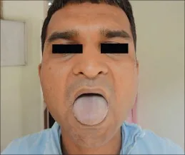
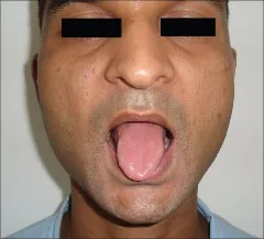
Central Cyanosis4 and Cyanosis Resolved4
Why Check: Central cyanosis indicates poor oxygenation in the lungs (specifically PaO2 <50mmHg given an Hb of 140-160g/L; PaO2 45mmHg if Hb 120 g/L); can occur in hypervolemic states e.g. pulmonary edema secondary to congestive heart failure or nephrotic syndrome. Images show tongue colouration changes before and after resolution of central cyanosis3.
Hypovolemia
Altered Mental Status/Confusion
AFMC level Pre-clerkship 3 · Clerkship 4
Say: I will briefly assess how you are doing cognitively.
Do: Can you tell me where you are right now? What kind of place is this? Do you know what day it is? What month? Year? What is your name? Do you know my name?
Report: You are alert and oriented to person, place and time (Alert and Oriented x3)
Why Check: Altered Mental Status can be due to cerebral hypoxia from ↓ volume and cardiac output. It is a worrying sign of shock.
Sunken Eyes
Peds - [1, 3]
Say: I will now inspect the eyes to look for sunken-ness
Do: Look for signs of sunken eyes on inspection
Report: Eyes do not appear sunken on inspection
Why Check: Important sign for hypovolemia, especially in pediatrics. The lack of sunken eyes slightly decreases the likelihood of hypovolemia (LR- 0.5) in adult patients. In pediatric patients, sunken eyes mildly increase the likelihood of dehydration (LR+ 1.7, LR- 0.49)
Dry Mucous Membranes
Gen - [3, 4] Peds - [1, 3]
Say: I will now inspect the mucous membranes. Can you briefly open the mouth and tilt the head back slightly?
Do: Assess for dryness of mucous membranes of nose and mouth (lips, inner cheeks, tongue)
Report: Mucous membranes appear moist.
Why Check: In hypovolemia, might note chapped lips and dry mouth. Lack of dry mucus membranes slightly decreases the likelihood of hypovolemia (LR- 0.3) in adult patients. In pediatric patients, dry mucous membranes mildly increase the likelihood of dehydration (LR+ 1.7, LR- 0.41)
Longitudinal Tongue Furrows
Say: I will now inspect the tongue. Can you briefly open the mouth and stick out the tongue?
Do: Look for longitudinal furrows
Report: Tongue appears absent of longitudinal furrows
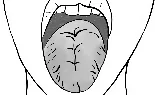
Why Check: Important sign for hypovolemic state. Lack of longitudinal tongue furrows slightly decreases the likelihood of hypovolemia (LR- 0.3) in adult patients.
Hyper- and Hypo-volemia
Jugular Venous Pressure (JVP) – important for both hyper- and hypo-volemia
JVP - [4, 4] JVP Waveform- [2, 3]
Also seen Ch 6 – Cardiac Exam – MF1
Say: Please lay back on the exam table and tilt the head all the way to the left.
How do you differentiate the JVP from the Carotid Pulse?
| JVP | Carotid |
|---|---|
| Occludable and bounces back after occlusion | Non-occludable |
| Non-palpable | Palpable |
| Biphasic | 1-1 Pulse to Heartbeat |
| Decreases: with elevated position and with inspiration | No change with position or inspiration |
| Increases: with Abdomino-Jugular Reflux (See below) | No change with AJR maneuver |
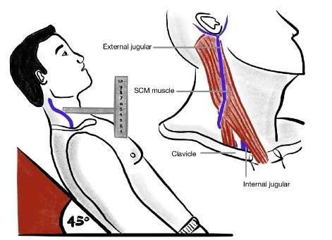
Do:
- Prop up head of exam table to 30-45°. Stand at right side. Look for JVP between 2 heads of sternocleidomastoid (i.e. sternal head medially, clavicular head laterally), or look lateral to sternal head of sternocleidomastoid.
- Once you have located the JVP, measure by placing ruler on sternal angle facing vertically
- Make a right-angle triangle going from the apex of the JVP to the ruler.
- The height on the ruler is the measured JVP.
Note: on OSCE, estimate using your fingers, < 3 finger breadths is normal – not expected to use ruler.
What is Normal: 3-4 cm Above Sternal Angle (ASA)
Report: JVP was located and is < 5 cm above the sternal angle
Why Check: JVP is the most common and perhaps most important sign to assess intravascular volume status of patients (termed “wet” if the patient is volume overloaded and the JVP is elevated, or “dry” if the patient is volume depleted and the JVP is flat). In general, the JVP will be flat in hypovolemic and distributive shock and elevated in cardiogenic and obstructive shock.
The JVP is an indirect measure of right atrial pressure via visualization of the internal jugular vein. The Central Venous Pressure (cm H2O) can be estimated from the JVP by adding 5cm to the measured value, since right atrium is approximately 5cm below the sternal angle. In the appropriate clinical context (e.g. dyspneic patients in Emergency Department), an elevated JVP increases the likelihood of suspected Congestive Heart Failure (LR+ 5.1; LR- 0.66) If the JVP increases with inspiration, this is called Kussmaul’s Sign and may be a sign of severe pericarditis or right-sided heart failure. Beck’s Triad (for Acute Cardiac Tamponade) includes distended neck veins, low Arterial Blood Pressure, and Muffled Heart Sounds – as a cause of Obstructive Shock.
Palpation
Hypervolemia
Edema
AFMC level Pre-clerkship 4 · Clerkship 4
(Also seen in Ch 6 – Cardiac Exam and Ch 8 – Peripheral Vascular Exam)
Say: I will palpate for leg swelling (Sacral edema if patient immobile). Please tell me if you are in discomfort.
Do: Press firmly with pads of your thumbs on the dorsal surface of patient’s leg, just above the ankle at bony prominence. Hold for 10s, then release. Look for pitting edema – skin does not bounce back immediately (i.e. pits). How high does it go on the leg? Is it pitting (transudate) or non-pitting (exudate)?
Look at patient for signs of discomfort and stop exam signs of distress.
Grading: 0 = none; 1+ = 2mm depression; 2+ = 4mm depression; 3+ = 6mm depression; 4+ = 8mm depression
Report: I note (grade) pitting edema bilaterally, up to the level of (location e.g. ankles).
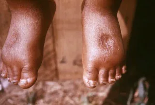
Pedal Edema5
Why Check: Edema is excess interstitial fluid accumulation causing swelling. Normally, this fluid returns through lymphatic drainage. Pitting edema = transudate (non-protein filled) – sign of increased hydrostatic pressure (e.g. congestive heart failure, decreased venous return) or decreased oncotic pressure (e.g. nephrotic syndrome, hypoalbuminemia). Dependent areas (e.g. legs if patient upright/mobile, sacrum if patient supine/bedbound) will become edematous with increased hydrostatic pressure or decreased oncotic pressure. Non-dependent areas (e.g. periorbital) will become edematous with decreased oncotic pressures (e.g. nephrotic syndrome).
Note: Non-pitting edema = exudate (protein) filled, or lymphatic drainage issue (e.g. lymphedema).
Hypovolemia
Decreased Skin-Turgor
Gen – [3, 4] Peds – [1, 3]
Say: I will palpate for skin turgor at the skin just below the clavicle.
Do: Pinch the skin and look for it recoil back into position
What is Normal: < 2 seconds is normal, > 2 seconds is poor skin turgor
Report: Skin turgor recoil occurs within 2 seconds, which is normal.
Why Check: Important sign for hypovolemia state. In pediatric patients, decreased skin turgor mildly increases the likelihood of dehydration (LR+ 2.5, LR- 0.66)
- Note: Limited utility in elderly patients – generally have physiologically decreased skin turgor.
Dry axilla
AFMC level Pre-clerkship 3 · Clerkship 4
Say: I will palpate the armpits to assess for signs of sweat.
Do: Using a tissue, place it under patient’s axilla for a few seconds, then withdraw and assess
Report: No signs of axillary dryness on palpation.
Why Check: In hypovolemia, the axilla will be dry as patients physiologically try to retain fluids. Dry axilla mildly increases the likelihood of hypovolemia (LR+ 2.8)
Capillary Refill Time
Gen – [4, 4] Peds – [1, 3]
Say: I will palpate for circulation in the fingers and toes. Can you raise one arm above the level of the heart?
Do: Press down on the nail bed until it turns colourless. If unable to see nail bed (e.g. with nail polish present), can press on pad of finger/toe as a backup. Record time when colour returns to the nail.
Normal Values: 2s for children and adult men, 3s for women, 4s for elderly; evidence suggests >5s for moderate-severe peripheral arterial disease.
Report: I note normal capillary refill time.
Why Check: Volume status sign, but also used to assess moderate-severe peripheral arterial disease. Prolonged capillary refill moderately increases the likelihood of hypovolemia (LR+ 6.9, LR- 0.7).
Hyper- and Hypo-volemia
Abdomino-jugular Reflux (AJR)
AFMC level Pre-clerkship 2 · Clerkship 3
(Hyper- and Hypo-volemia)
Say: Please lay back on the exam table and tilt the head to the left. I’m going to press on the upper abdomen and monitor the veins in the neck.
Do: Press firmly over right upper quadrant or centre of abdomen for 10-15 seconds. Observe the JVP. If JVP does rise significantly (≥4cm), observe if JVP returns to baseline while applying pressure. Sustained elevation of JVP beyond 10 seconds is abnormal. This can be confirmed by suddenly releasing abdominal pressure and seeing a return to baseline (≥4cm fall).
What is Normal: In an otherwise healthy individual, the JVP remains constant or temporarily rises for 1-2 heartbeats before returning to normal.
Report: Abdominojugular Reflux lasted less than 10 seconds, which is normal.
Why Check: An alternative test for measuring JVP, method to assess right ventricular failure. This is also useful if you are having trouble seeing or differentiating the JVP from carotid.
Central & Peripheral Pulses
AFMC level Pre-clerkship 1 · Clerkship 2
(Hyper- and Hypo-volemia)
See Ch 6 – Cardiac Exam and Ch 8 – Peripheral Vascular Exam
Say: I will palpate the central pulses in the neck, arms and groin.
Do: Palpate for pulses bilaterally. For peripheral pulses, you can palpate the most distal pulse. If it is non-palpable, move more proximally. Should be symmetrical, regular rate/rhythm and no delay between them.
Where to Palpate – Lower Extremities:
- Dorsalis Pedis: Dorsal side of foot, just lateral to extensor tendon of big toe
- Posterior Tibial: Posterior and inferior to medial malleolus = bony prominence on side of ankle
- Popliteal: Behind knees, use 2 hands
Where to Palpate – Upper Extremities:
- Radial: Along radius bone just proximal to wrist on palmar side of hand
- Brachial: Medial to biceps tendon and 2-3cm above antecubital fossa
Where to Palpate – Central Pulses:
- Carotid: Medial to sternocleidomastoid muscle, roughly at level of cricoid cartilage
- Abdominal Aorta: Slightly left and superior to umbilicus
- Femoral: Midway between ASIS and Symphysis pubis
Note: Auscultate for carotid bruits before palpating carotid pulses using bell of the stethoscope (see Chapter 6 – Cardiac Exam for details)
Grading: 0 = Non-palpable; 1 = Barely palpable; 2 = Easily Palpable (Normal); 3 = Bounding; 4 = Aneurysmal.
Report: The central and most distal peripheral pulses are symmetrical with regular rate and rhythm
Why Check: Pulses will feel bounding in hypervolemic state, and barely/non-palpable in hypovolemic state. In pediatric patients, weak pulses slightly increase likelihood of dehydration (LR+ 3.1-7.2). You can also assess for Pulses alternans (two strong systolic pulses with mid-systolic dip – suggests LV dysfunction or Cardiac tamponade) or Irregularly irregular rhythm (Atrial fibrillation).
Modification for Patients with Obesity: Use handheld Doppler Ultrasound to locate pulses if difficult to appreciate by palpation. In this population, a doppler may be more sensitive than palpation.
Temperature to Touch - Extremities
AFMC level Pre-clerkship 1 · Clerkship 2
(Hyper- and Hypo-volemia)
Say: I will palpate the top of each foot to check temperature on each side.
Do: Feel the dorsum of each foot with back of hand – assess whether it is cold to touch, or warm to touch. Migrate proximally on each side and compare temperature between each.
Report: I note no temperature deviation at the dorsal surface of the feet.
Why Check: In the context of a volume status exam, Temperature to touch is a common way to describe various shock states. Patients are often described as “cool and clammy” or “warm and flushed”. In particular, distributive shock states (due to decreased systemic vascular resistance) cause warm and flushed extremities, whereas hypovolemic, cardiogenic, and obstructive shock states (due to low cardiac output) cause cool and clammy extremities.
- Recall: In context of peripheral vascular exam (Ch 8 – Peripheral Vascular Exam), temperature can help differentiate arterial deficiency (cold feet) vs. venous deficiency (warm feet).
Auscultation
Listening to Breathing
General- [3, 4] Stridor - [4, 4] Pleural Rub – [1, 4] Stridor (Peds) - [1, 3] Wheeze (Infant) - [1, 2]
(Also seen in Ch 5 – Resp Exam)
Say: I will now listen to the chest and back – breathe in and out each time you feel the stethoscope. Please pause if you become light-headed.
Setup: Patient crosses arms to displace scapulae.
Do: Hold stethoscope still when listening (don’t want to only hear rustling of moving it around).
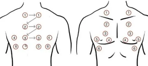
Report: I note air entry bilaterally with vesicular breath sounds and no adventitious sounds.
In the context of a Volume status exam, primarily listening for signs of hypervolemia e.g. diminished breath sounds which greatly increases likelihood of Pleural Fluid (LR+ 18.6), fine crackles (e.g. in congestive heart failure) or coarse crackles (e.g. with pulmonary edema) particularly in dependent areas (e.g. at lung bases).
What is Normal? Normal auscultation depends on listening location
- (1) Trachea: Bronchial – harsh, high-pitched, heard through full inspiration and expiration
- (2) Sternum: Bronchovesicular – less harsh, heard through full inspiration, short and softer on expiration
- (3) Everywhere else: Vesicular - gentle, low-pitched, quiet on inspiration and nearly silent on expiration
Why Check: Assessing for Adventitious (Abnormal) Sounds, including:
| Sound | Respiratory Phase | Description | Significance |
|---|---|---|---|
| Fine Crackles | Inspiration | Discontinuous, soft, high-pitched, more per breath, popping sound. | Distal lower airway process (e.g. pulmonary fibrosis, congestive heart failure) |
| Coarse Crackles | Inspiration alone; or Inspiration + early expiration | Discontinuous, loud, low-pitched, fewer per breath, bubbling sound. | Proximal lower airway process (e.g. pneumonia, pulmonary edema, adult respiratory distress syndrome) |
| Stridor | Inspiration | Loud, musical sound, constant pitch, louder over neck. | Upper airway obstruction (e.g. croup, foreign body aspiration, epiglottitis, bacterial tracheitis, retropharyngeal abscess, peritonsillar abscess) |
| Pleural Rub | Expiration alone; or Expiration + Inspiration | Loud, grating or rubbing sound; can be crackling. | Pleural inflammation (e.g. pleural effusion, pulmonary embolism, pleuritis) |
| Wheeze | Expiration alone; or Expiration + Inspiration | Continuous, high-pitched, musical sound. | ↓ Distal lower airway diameter (e.g. asthma, chronic obstructive pulmonary disease) |
| Rhonchus | Expiration alone; or Expiration + Inspiration | Continuous, low-pitched, coarse sound; may clear after cough. | ↓ Proximal lower airway diameter by mucous secretions, lesions or foreign bodies (e.g. pneumonia, cystic fibrosis) |
- Bronchial breath sounds over lung (normal over trachea) points to consolidation
- Early Crackles increases likelihood of suspected obstructive disease (LR+ 14.6), and particularly for severe obstruction in patients with chronic airflow limitation (LR+ 20.8; LR- 0.1)
- Stridor: May be earliest sign of airway compromise.
- Note - Pediatrics: Wheezing in Infants: Respiratory exam of wheezing in infants shows good inter-rater reliability, but respiratory status overall is most reliable.6
- Issue: Adventitious sounds on auscultation is “the most ambiguous and confusing nomenclature in all of physical diagnosis” where clinicians use diverse terms to describe similar sounds6.
Cardiac Precordial Auscultation
S3, S4- [1, 3] Murmur [1, 3] Abnormal S1, S2 [1, 1] Position [1, 4]
(Also seen in Ch 6 – Cardiac Exam)
Say: I will auscultate the chest for heart sounds. Please breathe normally as I listen.
Do: Listen carefully with the diaphragm of the stethoscope at each valve region (Aortic, Pulmonic, Tricuspid, and Mitral). Listen for abnormal heart sounds or murmurs.
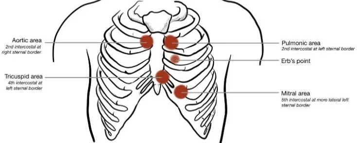
Report: I note a normal S1 and S2. There are no extra heart sounds or murmurs present.
What Is Normal? S1 and S2, regular rate and rhythm, with no extra heart sounds and no murmurs
- S1 = loudest at Tricuspid & Mitral valves (re atrioventricular valves closing at start of systole)
- S2 = loudest at Aortic & Pulmonic valves (re semilunar valves closing at end of systole). Physiologic splitting of S2 normal on inspiration due to ↑ -ve intrathoracic pressure – best heard in sitting position.
Abnormal Findings – Abnormal S1 and S2 and Associated Conditions:
| Characteristic | S1 | S2 |
|---|---|---|
| Loud | Mitral Valve Prolapse (MVP); Mitral Stenosis (MS); Left Atrial Myxoma; Tachycardia | Systemic hypertension (loud A2) or Pulmonary hypertension (loud P2) |
| Faint | Mitral Regurgitation; Left Bundle Branch Block (LBBB); Long PR interval (1st degree AV block); Acute Aortic Regurgitation (AR) | Aortic Stenosis (LR+ 7.5, LR- 0.5) |
| Variable | AV Dissociation (LR+ 24.4, LR- 0.4); Atrial Fibrillation | N/A |
| Wide Split | Right Bundle Branch Block (RBBB); Right Atrial Myxoma | RBBB; Pulmonic Stenosis; Mitral Regurgitation; Ventricular Septal Defect (VSD); Right Ventricular Failure |
| Paradoxical Split | N/A | LBBB; Severe Aortic Stenosis; Wolff-Parkinson-White (WPW); LV Dysfunction |
| Fixed Split | N/A | Atrial Septal Defect (ASD) |
Abnormal Findings – Extra heart sounds: Best heard in left lateral decubitus position.
- S3: Heard just after S2, with rapid and early diastolic filling of ventricles, “Sloshing in” (e.g. volume overload). In the appropriate clinical context (e.g. dyspneic patients in Emergency Department), hearing an S3 increases the likelihood of Congestive Heart Failure (LR+11, LR- 0.88).
- S4: Heard just before S1, with diastolic rapid late filling of ventricles, “A stiff wall” (e.g. pressure overload, Left Ventricular Hypertrophy).
- S3 + S4: Summation gallop
Abnormal Findings – Murmurs (represent Turbulent blood flow). Listen for Location (i.e. identify valve); Radiation (e.g. to carotids, left sternal border, etc.); Timing (e.g. Systolic vs. Diastolic); Configuration (e.g. Crescendo, Decrescendo, Crescendo-decrescendo, Plateau); Quality (e.g. high vs. low pitch; harsh, musical, blowing, rumbling, etc.); Change with maneuvers (e.g. with inspiration, Valsalva, squat); and Volume/Grade
- Systolic Murmur: Grades 1 (very faint); Grade 2 (soft, readily detectable); Grade 3 (easily detectable without thrill); Grade 4 (easily detectable with thrill); Grade 5 (very loud murmur audible with stethoscope placed lightly on chest); Grade 6 (extremely loud, audible with stethoscope off chest)
- Diastolic Grade: Grade 1 (barely audible); Grade 2 (audible, soft); Grade 3 (easily audible); Grade 4 (loud)
Common Murmur Description
Mitral Stenosis (MS)
- Pathophysiology: Mitral valve should be open in diastole – mitral stenosis causes incomplete opening of mitral valve in diastole
- Murmur: Diastolic murmur with Opening Snap best heard at cardiac apex in left lateral decubitus position. Worsened by ↑ venous return (squat/leg raise)
Aortic Regurgitation / Insufficiency (AR/AI)
- Pathophysiology: Aortic valve should be closed in diastole – aortic regurgitation causes incomplete closing of aortic valve in diastole
- Murmur: Diastolic murmur heard best at right base in sitting position. Worsened by ↑ venous return (squat/leg raise)
Aortic Stenosis (AS)
- Pathophysiology: Aortic valve should be open in systole – aortic stenosis causes incomplete opening of aortic valve in systole
- Murmur: Systolic murmur with crescendo-decrescendo pattern heard best at right base. Radiates to R clavicle and/or R carotid. Worsened by ↑ venous return (squat/leg raise)
Hypertrophic Cardiomyopathy (HCM)
- Pathophysiology: LV Outflow Tract Obstruction due to sarcomere gene mutations.
- Murmur: Systolic murmur with crescendo-decrescendo pattern heard best at cardiac apex and left lower sternal border. May radiate to axilla. Improved by ↑ venous return (squat/leg raise)
Mitral Regurgitation (MR)
- Pathophysiology: Mitral valve should be closed in systole – mitral regurgitation causes incomplete closing of mitral valve in systole.
- Murmur: Systolic murmur with holosystolic pattern heard best at cardiac apex. Radiates to axilla. Worsened by ↑ venous return (squat/leg raise)
Mitral Valve Prolapse (MVP)
- Pathophysiology: Mitral valve should be closed in systole, where thickened mitral valve leaflets in MVP cause imperfect closure of mitral valve in systole.
- Murmur: Systolic murmur similar to Mitral regurgitation, but improved by ↑ venous return (squat/leg raise)
Maneuvers to Accentuate/Differentiate Murmurs:
| Maneuver | RV Preload | LV Preload | LV Afterload | Effect on Murmurs |
|---|---|---|---|---|
| Inspiration | ↑ | ↓ | ∅ | ↑ R-sided murmurs, ↓ L-sided murmurs (except HCM) |
| Valsalva (Strain Phase)/ Abrupt Stand | ↓ | ↓ | ↓ | ↑ MVP/HCM, ↓ MS/MR/AS/AR/VSD |
| Squatting/Leg raise | ↑ | ↑ | ∅/↑ | ↓ MVP/HCM, ↑ MS/MR/AS/AR/VSD |
| Hand Grip (for 20-30s) | ∅ | ∅ | ↑ | ↓ MVP/HCM; ↑ MR/AR/VSD; ↓ MS/AS |
Note on Pediatric Cardiac Auscultation: Not all pediatric murmurs are pathologic; pathology more likely if murmur is systolic grade 3+; holosystolic; associated systolic click; diastolic; worse on standing; or harsh.
Helpful Stethopedia Videos to Review
- Video 1 – Renal and Volume Status Exam OSCE Style (Link: https://videopress.com/v/ej80D4WR)
Part 5: Relevant Likelihood Ratios – from Rational Clinical Examination JAMA series
Clinical Question 1: Is this patient Hypovolemic (Adult)?7
Note: The following reflect diagnostic accuracy of physical signs for hypovolemia not due to blood loss.
| Procedure | Sensitivity | Specificity | LR+ (95% CI) | LR- (95% CI) |
|---|---|---|---|---|
| Capillary Refill Time Elevated | 0.34 | 0.95 | 6.9 (3.2-15) (about +30%) | 0.7 (0.5-0.9) (about −15%) |
| Sunken Eyes | 0.62 | 0.82 | 3.4 (1.0-12) (no change: 95% CI includes 1) | 0.5 (0.3-0.7) (about −15%) |
| Speech not clear or expressive | 0.56 | 0.82 | 3.1 (0.9-11) (no change: 95% CI includes 1) | 0.5 (0.4-0.8) (about −15%) |
| Dry Axilla | 0.50 | 0.82 | 2.8 (1.4-5.4) (about +15%) | 0.6 (0.4-1.0) (no change: 95% CI includes 1) |
| Upper/Lower Extremity Weakness Present | 0.43 | 0.82 | 2.3 (0.6-8.6) (no change: 95% CI includes 1) | 0.7 (0.5-1.0) (no change: 95% CI includes 1) |
| Tongue Dry | 0.59 | 0.73 | 2.1 (0.8-5.8) (no change: 95% CI includes 1) | 0.6 (0.3-1.0) (no change: 95% CI includes 1) |
| Confusion Present | 0.57 | 0.73 | 2.1 (0.8-5.7) (no change: 95% CI includes 1) | 0.6 (0.4-1.0) (no change: 95% CI includes 1) |
| Dry mucous membranes of mouth and nose | 0.85 | 0.58 | 2.0 (1.0-4.0) (no change: 95% CI includes 1) | 0.3 (0.1-0.6) (about −30%) |
| Longitudinal Furrows on Tongue | 0.85 | 0.58 | 2.0 (1.0-4.0) (no change: 95% CI includes 1) | 0.3 (0.1-0.6) (about −30%) |
| Postural Vitals-Pulse Increment > 30 bpm | 0.43 | 0.75 | 1.7 (0.7-4.0) (no change: 95% CI includes 1) | 0.8 (0.5-1.3) (no change: 95% CI includes 1) |
| Postural Vitals –SBP decline > 20 mmHg | 0.29 | 0.81 | 1.5 (0.5-4.6) (no change: 95% CI includes 1) | 0.9 (0.6-1.3) (no change: 95% CI includes 1) |
Clinical Question 2: Is this child Dehydrated? (5% Dehydration)8
| Procedure | Sensitivity | Specificity | LR+ (95% CI) | LR- (95% CI) |
|---|---|---|---|---|
| Prolonged Capillary Refill | 0.60 (0.29-0.91) | 0.85 (0.72-0.98) | 4.1 (1.7-9.8) (about +15%) | 0.57 (0.39-0.82) (about −15%) |
| Weak Pulse | 0.04-0.25 | 0.86-1.0 | 3.1-7.2 (about +15%) | 0.66-0.96 (about −15%) |
| Abnormal Skin Turgor | 0.58 (0.40-0.75) | 0.76 (0.59-0.93) | 2.5 (1.5-4.2) (about +15%) | 0.66 (0.57-0.75) (about −15%) |
| Absent Tears | 0.63 (0.42-0.84) | 0.68 (0.43-0.94) | 2.3 (0.9-5.8) (no change: 95% CI includes 1) | 0.54 (0.26-1.1) (no change: 95% CI includes 1) |
| Abnormal Respiratory Pattern | 0.43 (0.31-0.55) | 0.79 (0.72-0.86) | 2.0 (1.5-2.7) (about +15%) | 0.76 (0.62-0.88) (about −15%) |
| Poor Overall Appearance | 0.80 (0.57-1.0) | 0.45 (-0.1-1.0) | 1.9 (0.97-3.8) (no change: 95% CI includes 1) | 0.46 (0.34-0.61) (about −30%) |
| Dry Mucous Membranes | 0.86 (0.80-0.92) | 0.44 (0.13-0.74) | 1.7 (1.1-2.6) (about +15%) | 0.41 (0.21-0.79) (about −30%) |
| Sunken Eyes | 0.75 (0.62-0.88) | 0.52 (0.22-0.81) | 1.7 (1.1-2.5) (about +15%) | 0.49 (0.38-0.63) (about −30%) |
| Cool Extremity | 0.10-0.11 | 0.93-1.0 | 1.5-19 (about +15%) | 0.89-0.97 (about −15%) |
| Increased Heart Rate | 0.52 (0.44-0.60) | 0.58 (0.33-0.82) | 1.3 (0.8-2.0) (no change: 95% CI includes 1) | 0.82 (0.64-1.0) (no change: 95% CI includes 1) |
| Sunken Fontanelle | 0.49 (0.37-0.60) | 0.54 (0.22-0.87) | 0.9 (0.6-1.3) (no change: 95% CI includes 1) | 1.12 (0.82-1.5) (no change: 95% CI includes 1) |
References
-
Evans L, et al. Surviving Sepsis Campaign: international guidelines for management of sepsis and septic shock 2021. Crit Care Med. 2021;49(11):e1063-e1143. ↩
-
Recommendations. Choosing Wisely Canada. Accessed August 1, 2022. https://choosingwiselycanada.org/recommendations/#list ↩
-
McDonald M, et al. CCS/CHFS heart failure guidelines update: defining a new pharmacologic standard of care for heart failure with reduced ejection fraction. Can J Cardiol. 2021;37(4):531-546. ↩ ↩2
-
Das J, Katyal A, Naunwaar D. Dapsone-induced methemoglobinemia in a patient of leprosy. Indian J Dermatol. 2015;60(1):108. ↩ ↩2
-
Centers for Disease Control and Prevention. Accessed February 21, 2023. https://phil.cdc.gov/Details.aspx?pid=16536 ↩
-
McGee S. Evidence Based Physical Diagnosis. 4th ed. Elsevier; 2017:268. ↩ ↩2
-
Is this adult patient hypovolemic. In: Rational Clinical Examination Series. Chapter 24. 2009:320. ↩
-
Is this child dehydrated. In: Rational Clinical Examination Series. Chapter 25. 2009:334. ↩