Medical Foundations 3
Chapter 21Clinical Breast Examination
Part 1: Focused History – Based on Chief Complaint (Begin with Open-Ended Questions)
HPI, Associated Symptoms
Breast Lump
- Location and when/how it was found?
- Shape and size?
- Consistency – Hard vs. Soft, Nodular vs. Uniform, Mobile vs. Fixed?
- Contour – Well-Defined vs. Ill-Defined?
- Tender, Red or Warm?
- Correlated changes in menstrual cycle?
Affected Breast Appearance
- Skin or nipple retraction? (re: Cooper’s ligaments)
- Dimpling?
Skin Changes
- Eczema, rash, ulceration or skin breakdown on breast?
- Injury or fat necrosis?
Nipple Discharge
- Bloody, milky, clear or purulent? Bilateral or unilateral?
Constitutional Symptoms
- Fever, chills, night sweats, weight loss, anorexia or asthenia (weakness)
Past Medical History (PMHx)
- Breast Cancer Screening: Last mammography, breast ultrasound
- Current Cancer Care Ontario Recommendations (e.g. Breast Cancer Screening)
- Pro Comp Connection: Cancer screening is an important opportunity for patient-centred shared decision making (e.g. using patient-centred decision aid tools)
- Previous Investigations: Mammography, Breast Biopsy and Cytologic Studies (cyst aspiration, nipple discharge)
- Obstetrical/Gynecological History:
- Previous breast malignancy or dysplasia; Previous breast lumps, fibrocystic changes
- Age of menarche (earlier menarche ↑ breast cancer risk)
- Age at first pregnancy (later first pregnancy ↑ breast cancer risk)
- Past pregnancies (Nulliparity ↑ breast cancer risk), Prior use of any fertility treatment?
- Age of menopause (later menopause ↑ breast cancer risk – median age of menopause is 51.4 years)
- Breastfeeding (↓ breast cancer risk)
- Trauma to breast
- Oncological history: breast cancer in contralateral breast, endometrial cancer, ovarian cancer, colorectal cancer, or radiation therapy (e.g. ionizing radiation for Hodgkin’s Lymphoma ↑ breast cancer risk.
Past Surgical History (PS Hx)
- Prior lumpectomies, mastectomy; breast reduction surgeries; breast implant procedures & complications
Family Medical History (FMHx)
- Breast Cancer (especially first-degree relatives, or multiple family members on a side of family)
- Ovarian, Endometrial and Uterine Cancer (especially 1st degree relatives, or multiple family members on a side of family)
- BRCA-1 and BRCA-2 mutations confer risk of cancer: Only way for your screening test to mean anything above baseline population risk is to have a close relative tested for BRCA-1/BRCA-2 who also has diagnosed breast cancer.
- Lynch Syndrome (Autosomal dominant impairing DNA mismatch repair - condition with ↑↑ risk of several cancers)
- Any Relatives Diagnosed with Cancer at Early Age
- Black Patients at ↑ risk of triple-negative breast cancer (i.e. ER- PR-, HER2-; more difficult to treat)
Social Hx
- Smoking and Alcohol intake → ↑ breast cancer risk
- Others: Illicit Drug use; Nutrition, Family & Social Support Network; Physical activity
Medications Hx & Allergies
- Immunosuppressive medications → Increased malignancy risks
- Combined Estrogen-Progestogen Hormone Replacement Therapy (HRT)2: ↑ Breast and Ovarian cancer risk with long-term use (≥5 years); taking Estrogen without progestogen component ↑ risk of endometrial cancer.
- Combined Oral Estrogen-Progestogen Contraceptives (OCP)2: ↑ Breast, cervical and liver cancer risk while on treatment; cessation of OCP use ↓ cancer risk in dose-dependent manner, with no excess risk by 10 years off treatment.
Part 2: Pertinent Notes about the Physical Exam
- Key Idea: It is essential to conduct sensitive exams in a patient-centred, trauma-informed way before, during, and after exams. This includes close attention to privacy, consent, draping, verbal communication and non-verbal communication. For more information, see Ch 4 – Vital Signs and Introduction to Patient-Centred Physical Exams – MF1.
- Third-Party Attendance at Intimate Examinations (See CPSO Policy – Boundary Violations)3: Physicians must give patients the option of having a third-party present (e.g. Support Person, or Chaperone) during an intimate examination, including bringing their own third party if the physician does not have one (See Ch 4 – Vital Signs – MF1 for more info).
Part 3: Specific Maneuvers in the Physical Exam – Say, Setup, Do, Report
Inspection
Localized Differences
AFMC level Pre-clerkship 2 · Clerkship 3
Say: I will inspect for localized differences between the breasts such as lumps, skin dimples and differences.
Setup: Patient should be sitting with both arms at sides, hands on knees initially.
Say: Could you lie back on the exam table, with one arm resting under your head?
Do: Inspect the skin for changes, as well as the nipple for discharge. Check the skin for Peau d’orange (Orange Peel Skin), Dimples (Ask patient to lift arms or press on hips re: tensing pectoral muscles, pulls on Cooper’s ligaments to reveal dimpling), Retracted Nipples, or Dermatitis of Areola. If you note any discharge, comment on Amount, Consistency, Colour and Odor. Also inspect for redness (mastitis) and for breast abscess.
Say: Can you switch the arm resting under your head?
Do: Repeat inspection for same features on other breast
Report: USE CLOCK METHOD TO REPORT FINDING (X minutes from nipple to finding). I note no discharge, redness, skin changes, dimpling, nipple inversion or abscesses.
Why Check: No longer part of screening for breast cancer (no difference in outcomes over & above mammogram). Still a part of clinical encounters (e.g. for symptomatic patients). Now the focus is on palpation. While you are palpating the breast, you can continue to inspect for the findings outlined above as well.
- Malignant: Bloody discharge may represent intraductal papilloma (benign papillary tumor in lining of breast duct) or breast cancer (e.g. ductal carcinoma in situ, invasive ductal carcinoma). Skin erythema, retraction or dimpling, peau d’orange, nipple inversion may all be signs of locally advanced breast cancer. Peau d’orange with tenderness, burning and blood-tinged nipple discharge may be seen in inflammatory breast cancer. Erythematous/scaly/vesicular/ulcerated rash that begins at the nipple and spreads to areola may represent Paget disease of the breast.
- Non-malignant: Clear or slightly milky nipple discharge may represent fibrocystic breast changes (most common benign lesion). Milky or bloody nipple discharge can also represent mastitis if present with signs of inflammation (e.g. swelling, erythema, warmth). Skin retraction, erythema and bruising may represent fat necrosis (benign). A fluctuant, tender, palpable mass with red streaks around the mass and constitutional symptoms may represent a breast abscess. Eczema of the breast (benign) may present with an eczematous rash with poor margins and pruritis and may become complicated by secondary cellulitis. In lactating patients, a plugged duct may present with a tender and painful lump without systemic findings.
- Presenting with new symptoms of breast cancer (e.g. concerning nipple discharge, new nipple retraction, sin changes of nipple or breast, asymmetric thickening/nodularity) warrants further imaging2:
- Patient <30 years old and/or breastfeeding or pregnant → Ultrasound (re. density of breast tissue)
- Patient 30-40 years old → Ultrasound ± Mammogram ± Galactography (in appropriate context)
- Patient >40 years old → Ultrasound and Mammogram ± Galactography (in appropriate context)
Modification for Patients who are Transgender:
- Transmasculine Patients (FTM): Transmasculine patients may use a binder to flatten their chest. They may not use the word “breast” and may instead refer to their chest or use another word of their choice. If they have had top surgery, you may see evidence of a scar and/or chest contouring.
- Transfeminine Patients (MTF): Patients on feminizing hormone therapy may have some breast tissue development. If they have had top surgery (aka breast augmentation), you may notice breast implants and/or nipple reconstruction on exam, and you may see evidence of scars from the procedure.
Tanner Staging (Adolescent Females) – Breast Development
Say: I will inspect the breasts for adolescent development.
Do: Inspect the breasts according to Tanner Staging.
What is Normal Breast Tanner Staging?
- Stage 1 – No glandular tissue, areola following skin contours of breast (Age 10 and younger)
- Stage 2 – Breast bud forms, small area of glandular tissue around it; areola widens (Age 10-11.5)
- Stage 3 – Breast becomes more elevated, extends beyond areola which continues to be in contour with rest of the surrounding breast (Age 11.5-13)
- Stage 4 – Increased size and elevation; areola and papilla form secondary mound projection from contour of surrounding breast (Age 13-15)
- Stage 5 – Final adult size; areola in contour with breast, projecting central papilla (Age 15+)
Report: Based on tanner staging of breasts, patient is in Tanner Stage 4.
Why Check: Important tool for measuring development based on primary and secondary sex characteristics – for Tanner Staging of Pubic Hair, see Chapter 19 – Sensitive Pelvic Examinations; normal progression is breast development (thelarche) → pubic hair development (pubarche) → growth spurt → menarche.
Modification for Transfeminine Patients (MTF): Patients on feminizing hormone therapy may have some breast tissue development. If they have had top surgery (aka breast augmentation), you may notice breast implants and/or nipple reconstruction on exam, and you may see evidence of scars from the procedure.
Palpation
Lumps Assessment
AFMC level Pre-clerkship 2 · Clerkship 3
Say: I will now palpate the breast. Can you roll the pelvis to the opposite side, bring the shoulder back and make it flat with the exam table, and place the hand on your forehead in a “drama queen” pose?
- Once palpation reaches the midclavicular line, ask patient to change positions to lay flat on the exam table, and have their elbow raised to the same level as their shoulder. This will shift breast tissue and make the medial half of the breast flat with the chest wall so you can palpate more medial structures
- Note: Your technique depends on breast size. If the breast is large and extends over the abdominal wall, you will need to have one hand underneath the section of the breast sitting on top of the abdominal wall, while your other hand is palpating the breast anteriorly.
Do: Breast palpation for lumps assessment should take ~ 3 minutes per breast.
-
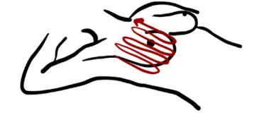
Breasts are bounded superiorly by the clavicle, inferiorly by the inframammary crease (“bra line”), medially by the sternum, and laterally by the mid-axillary line.
-
Palpate each breast using the Vertical Pattern technique (“Mowing the lawn”) with 3 pads of fingers with enough pressure you bring the skin with you as you palpate in circles (Light Pressure → Medium → Firm).
-
With patient in “drama queen” pose, start mid-axillary line, high near axilla, down to the inframammary crease (“bra line”), mowing lawn technique proceeding medially, palpating the lateral breast.
-
Once reaching Midclavicular line, have patient put hips flat and elbow raised to same level of the shoulder to shift structures so medial breast now at chest wall, and continue palpating.
-
Once reaching nipple, use “claw” technique. Note tenderness, pulsatility, warmth & colour. If you detect lesions, note size, shape, location, consistency and colour. Check for discharge from the nipple.
Report: I note no concerning lumps (if you do, use clock method re X minutes from nipple to finding).
Why Check: Looking for evidence of cysts or cancerous growths.
- Breast Implants: Placed deep to pectoral muscle; therefore, any lumps will be above it
- Cysts – Round lump (Mushroom)
- Cancerous – Multi-lobed (Broccoli)
- Presenting with new symptoms of breast cancer (e.g. palpable mass) warrants further imaging4:
- Patient <30 years old and/or breastfeeding or pregnant → Ultrasound (re. density of breast tissue)
- Patient 30-40 years old → Ultrasound ± Mammogram ± Galactography (in appropriate context)
- Patient >40 years old → Ultrasound and Mammogram ± Galactography (in appropriate context)
Lymph Nodes
AFMC level Pre-clerkship 3 · Clerkship 4
- Note: Important to include palpation of lymph nodes in the supraclavicular, Infraclavicular & axillary Lymph Node regions; for how to conduct a Lymph Node exam, see Chapter 11 – Lymph Node Exam –MF2
Why Check: Lymphadenopathy in these regions can be indicative of metastasis of breast cancer
Part 4: Relevant Likelihood Ratios – from Rational Clinical Examination JAMA series
Clinical Question 1: Does This Patient Have Breast Cancer? (Clinical Breast Examination)3
*Note: This article contains additional helpful images for a systematic approach to palpation in the clinical breast examination.
| Population | LR+ (95% CI) | LR- (95% CI) |
|---|---|---|
| Asymptomatic Patient (i.e. Screening Clinical Breast Exam) | 9.5 (8.9-10) (about +30%) | 0.66 (0.64-0.69) (about −15%) |
| Symptomatic Patient (e.g. pain, lump, nipple discharge, change in nipple or skin) | 2.5 (2.4-2.6) (about +15%) | 0.22 (0.20-0.25) (about −30%) |
References
-
Recommendations. Choosing Wisely Canada. Accessed August 1, 2022. https://choosingwiselycanada.org/recommendations/#list ↩
-
Cancer Care Ontario. Accessed June 21, 2022. https://www.cancercareontario.ca/sites/ccocancercare/files/assets/CCORiskFactorsReproductiveFemale.pdf ↩ ↩2 ↩3
-
Does this patient have breast cancer? The screening clinical breast examination: should it be done? How? – update. In: Rational Clinical Examination Series. Chapter 8. 2009:99-101. ↩ ↩2
-
Cancer Care Ontario. Accessed June 21, 2022. https://www.cancercareontario.ca/sites/ccocancercare/files/assets/CCOBreastScreeningPathwayMap.pdf ↩