Medical Foundations 4
Chapter 24Elbow Examination
Part 1: Focused History – Based on Chief Complaint (Begin with Open-Ended Questions)
HPI
Pain Assessment
- Onset:
- Acute onset of pain, disability immediate with injury (e.g. acute trauma, fracture) (see Mechanism of Injury below)
- Delayed onset of pain (e.g. ligamentous injury)
- Acute onset with no associated injury (e.g. septic arthritis)
- Insidious onset gradually worsening over time (e.g. overuse injury)
- Pediatric patients: Is the patient in an active growth spurt → increased risk of overuse injuries
- Provoking:
- Aggravating postures: Wrist flexion/extension; Forearm pronation/supination; Elbow flexion/extension
- Aggravating activities: Gripping (requires wrist stabilization); Lifting – distinguish everyday lifting activity (e.g. picking up a cup) vs. heavy activity (e.g. repeated weightlifting)
- Palliating:
- Relieving postures/activities: Neutral forearm pronation/supination; elbow flexion/extension; holding elbow close to body (e.g. in Radial Head Subluxation)
- Previous treatment: POLICE (Protect, Optimally Load, Ice, Compress, Elevate)? Physio? Analgesics?
- Rehab: Posture changes to activity; manual therapy; exercise (e.g. resistance, ROM); elbow brace, cast
- Investigations: Type (x-rays, Ultrasound, MRI, etc.) and results (if available)
- Quality: Deep ache, sharp vs. dull? – Ask patient what words they would use
- Neuropathic Pain Descriptors: Pins/needles; hot/burning/stinging; numb; electrical shocks; sensitivity to touch (e.g. clothing, bed sheets)
- Region: Poorly localized (e.g. Radial head subluxation) vs. Well-localized (e.g. Lateral epicondylitis)
- Elbow pain: Lateral vs. Medial vs. Posterior – helps organize differential diagnosis
- Radiation: For example, radiation along a peripheral nerve (e.g. Cubital Tunnel Syndrome)
- Severity: Immediately after injury, vs. subsequently. Has it improved over time with rest?
- Was patient able to continue their activity, e.g. after the sport completed, noticed pain got worse?
- Timing: Constant vs. Intermittent
- Recurrence: Assess episodes and pattern of recurrence of pain
- Associated Symptoms:
- Hearing/Feeling a “pop” – Acute tendon rupture (e.g. Medial collateral ligament rupture) or Avulsion Fracture (e.g. Avulsion Fracture of Medial Epicondyle – Pediatrics)
- Sensory Neuropathy e.g. paresthesias or numbness – assess distribution e.g. Peripheral nerve, Dermatomal or Spinal level (See Ch 31 – Screening Neuro Exam) – e.g. impaired sensation over ulnar nerve distribution in Cubital Tunnel Syndrome or Avulsion Fracture of Medial Epicondyle (pediatrics)
- Weakness: Weakness in specific motion may indicate a muscle tear; if concern for a neurologic cause (e.g. Ulnar nerve injury causing intrinsic hand muscle weakness), may need to do a screening neuro exam (See Ch 31 – Screening Neuro Exam)
Red Flags
- Pain: Pain at night/rest/unrelated to activity; Pain with passive movement; Poor Response to Treatment
- Constitutional Symptoms: Fever, night sweats, weight loss, anorexia
- Acute Compartment Syndrome (Can occur minutes-hours after acute injury, particularly closed fracture with compartments intact) → Surgical emergency (fasciotomy relieves compartment pressure)
- 6 Ps of compartment syndrome: pain, poikilothermia, paresthesia, paralysis, pulselessness, pallor.
- Pain: Out of proportion; not improving with analgesia; at rest; with passive muscle stretching
- LATE Findings of acute compartment syndrome: Pallor, paresthesia, paralysis, and pulselessness
- High Risk Facture of upper limb: Supracondylar Fracture (Pediatrics)
- Fullness or distension of involved compartment
- Inflammatory Arthritis:
- Intra-articular features: New onset joint swelling/effusion; Morning stiffness >30 mins (vs. <30 mins in Osteoarthritis); Improves during day and with activity (vs. worsens in osteoarthritis); distribution (Monoarthritis = 1, Oligoarthritis = 2-4, Polyarthritis = 5+ joints, symmetrical vs. asymmetrical)
- Extraarticular features: Psoriasis, Inflammatory Bowel Disease; Uveitis/Iritis; Photosensitivity rash; Raynaud’s Phenomenon; Urethritis; Diarrheal Illness
- Septic Arthritis: Acute atraumatic pain. Look for adjacent skin infection (vs. hematogenous seeding e.g. in a patient with sepsis), fevers and chills with a warm, erythematous, swollen joint and painful restricted range of motion in all directions.
- Neurovascular Damage: High-velocity injury; no pulses; hand weakness
- Non-Accidental Injury (Pediatrics): Delayed seeking medical attention. History not fitting with injury presentation. Unwitnessed injury. Bruises over soft tissue areas. Prior history of injuries or neglect. Types of fracture (e.g. metaphyseal)
Mechanism of Injury
- If injury occurred during an activity, ask open-ended questions about mechanism of injury e.g. “run me through what happened, play-by-play”. Try to visualize what happened. Assess low energy vs. high energy force injury and the forces involved (e.g. direct vs. indirect forces and their direction)
- If assessing an athlete, helpful to know/learn about their sport to understand common injuries and the demands of the position they play in the sport.
- Lateral epicondylitis (tennis elbow) can commonly occur in activities requiring gripping and wrist extension. Medial epicondylitis (golfer’s elbow) can commonly occur in overuse of wrist flexors.
Lateral Elbow Pain (most common site of elbow pain)
- Common Illness Presentations:
- Lateral Epicondylitis (Extensor Tendinopathy – “Tennis Elbow”): Localized elbow pain to lateral epicondyle and proximal forearm. Pain worsened by gripping or wrist extension. No pain in neck, upper arm or beyond the wrist. An often-insidious onset may be provoked by overuse (e.g. repeated gripping or wrist extension activity), or may be acute onset (e.g. lifting heavy object, or single-handed backhand in tennis)
- Radiohumeral Bursitis: Acute onset of pain over radiohumeral joint distal to lateral epicondyle, with maximal tenderness over anterolateral aspect of head of the radius and pain limiting range of motion (e.g. pronation/supination). No pain in neck, upper arm or beyond the wrist.
- Radial Head Subluxation (Pediatrics e.g. 1-4 years old – “Pulled Elbow”): Acute onset, poorly localized pain with “pull injury” – caregiver grabs child’s arm to prevent falling or pulling away; also occurs during play (e.g. being swung by forearms). Patient refuses to use the arm and may hold arm close to body with forearm pronated and elbow either fully extended or slightly flexed and has pain with supination.
- Can’t Miss Diagnosis Illness Presentations:
- Radial Head Fracture (most common elbow fracture): Acute onset lateral elbow pain with immediate swelling and limited forearm pronation/supination. Occurring due to Fall on Outstretched Hand (FOOSH; e.g., fall from height or bicycle). Can present with coronoid fracture (see below) and elbow dislocation (see below) – called the Terrible Triad.
- Pediatrics – Osteochondritis Dissecans of Capitellum (Repetitive compressive forces between radial head and capitellum): Gradual onset of lateral elbow pain worse with activity (e.g., throwing). In advanced cases, may present with symptoms of popping, locking or catching. Can’t miss diagnosis due to risk of avascular necrosis of anterolateral capitellum, with reduced range of motion, and potentially ↑ risk of osteoarthritis.
- Referred Pain (Cervical or Upper Thoracic Spine): Pain worse with prolonged postures (e.g., sitting)
Medial Elbow Pain
- Common Illness Presentations:
- Medial Epicondylitis (Flexor/Pronator Tendinopathy – “Golfer’s Elbow”): Localized elbow pain at or below medial epicondyle. Pain worsened by lifting. Often insidious onset provoked by overuse of wrist flexors.
- Medial Collateral Ligament Sprain: Localized elbow pain located just distal to medial epicondyle that is worse in late cocking/early acceleration phase of throwing. An often insidious, gradual onset pain may be provoked by overuse (e.g. excess valgus stress due to throwing), or acute onset pain (e.g. due to MCL rupture – if complete disruption with associated elbow instability, surgical repair required) where patient hears or feels a “pop” and unable to continue throwing.
- Cubital Tunnel Syndrome (ulnar nerve compression at elbow): Posteromedial elbow pain with paresthesias/numbness along ulnar nerve distribution (ulnar border of forearm, and D4-D5 of hand). May present with tenderness behind medial epicondyle (where nerve passes in a superficial groove between olecranon and medial epicondyle) or reproduction of symptoms with tapping over the nerve (positive Tinel’s sign). May occur due to acute trauma (e.g. nerve laceration, distal humerus fracture), nerve compression (e.g. prolonged elbow flexion, leaning on elbow), or elbow joint pathology (e.g. arthritis, synovitis, etc.).
- Can’t Miss Diagnosis Illness Presentations:
- Coronoid Fracture: Acute onset medial elbow pain with immediate swelling, may present with elbow instability. Occurring due to FOOSH (e.g. fall from height or bicycle), or with direct trauma to elbow. Can present with radial head/neck fracture (see above) and elbow dislocation (see below) – called the Terrible Triad.
- Avulsion Fracture of Medial Epicondyle (Pediatrics): Acute onset medial elbow pain over medial epicondyle, worse with resisted wrist flexion. Occurs due to Posterior elbow dislocation (with force transmitted to medial epicondyle through ulnar collateral ligament) or FOOSH. Patient may hear a sudden “pop” at elbow at time of injury. present with ecchymosis and generalized swelling (e.g. if elbow dislocated). Can’t miss diagnosis with risk of ulnar nerve dysfunction.
- Referred Pain (Cervical or Upper Thoracic Spine): Pain worse with prolonged postures (e.g., sitting)
Posterior Elbow Pain
- Common Illness Presentations:
- Olecranon Bursitis: Localized pain at posterior elbow. Pain may be acute onset (e.g. after trauma) or gradual onset (e.g. with repeated falls onto posterior elbow, or resting elbow on hard surfaces for prolonged periods). Look for signs of infection (requires immediate drainage, immobilization and antibiotics – risk of septic arthritis or osteomyelitis if left untreated)
- Can’t Miss Diagnosis Illness Presentations:
- Pediatrics – Supracondylar Fracture (Fracture of distal humerus just above elbow): Acute onset elbow pain with refusal to move elbow and immediate swelling. Occurring due to FOOSH (e.g. fall from height or bicycle), or with direct trauma to elbow. Can’t miss diagnosis with risk of neurovascular compromise (e.g. compartment syndrome, median/ulnar/ radial neurologic dysfunction) –an Orthopedic Emergency.
- Olecranon Fracture: Acute onset pain over olecranon area with immediate swelling and inability to extend elbow. Occurring due to FOOSH (e.g. fall from height or bicycle), or with direct trauma to elbow. Commonly occur with high energy injuries in pediatric patients, or low energy falls in elderly patients.
- Posterior Dislocation: Acute onset pain in posterior elbow with immediate swelling and obvious deformity. Occurring after fall or twisting injury. Can present with radial head/neck fracture (see above) and coronoid fracture (see above) – called the Terrible Triad. Can’t miss diagnosis with risk of neurovascular compromise (e.g. pulselessness) which requires urgent reduction of fracture and urgent surgical intervention if reduction unsuccessful in restoring neurovascular status.
Past Medical History (PMHx)
- Previous Elbow injuries
- History of Elbow Fractures – ↑ risk of elbow stiffness (e.g. loss of terminal extension).
- Recent localized tendon corticosteroid injection: ↑ risk of tendon rupture.
- Risk Factors for Septic Arthritis: Immunosuppression (including diabetes mellitus and HIV); Pre-existing joint disease (e.g. Rheumatoid arthritis: LR+ 2.5, LR- 0.45); Advanced Age; Skin infection (LR+ 2.8, LR- 0.76)
- Lab Markers for Septic Arthritis:
- Peripheral – CRP >100 mg/L (LR+ 1.6, LR- 0.44), WBC >10 (LR+ 1.4), ESR >30mm/h (LR+ 1.3)
- Synovial fluid – WBC >100 (LR+ 28, LR- 0.71), WBC >50 (LR+ 7.7, LR- 0.42), WBC >25 (LR+ 2.9, LR- 0.32), PMN cells ≥90% (LR+ 3.4, LR- 0.34)
- Lab Markers for Septic Arthritis:
- Bleeding Disorder History: ↑ risk of serious pathology on injury
- Risk Factors for Osteoarthritis: Age (>50 years); Female Gender; Obesity; Heavy manual labour; History of joint injury
- Psoriasis: Risk factor for Psoriatic Arthritis.
- Autoimmune disorders (↑ risk for Inflammatory Arthritis) – particularly Spondyloarthropathies e.g. Psoriatic arthritis, Uveitis, Ankylosing Spondylitis, Inflammatory Bowel Disease, Reactive Arthritis
- Risk Factors for Gout: Obesity, Hypertension, Dyslipidemia, Diabetes Mellitus, Chronic Kidney Disease
Past Surgical History (PS Hx)
- Arthroscopic surgery e.g. Medial Collateral Ligament reconstruction (“Tommy John Surgery”)
- Open-Reduction-Internal-Fixation, Pinning e.g. previous Radial Head Fracture etc.
- Ulnar nerve decompression e.g. for Cubital Tunnel Syndrome
- Palliative Radiation (e.g. for Painful Bony Metastasis in cancer)
- Risk Factors for Septic Arthritis: Recent joint surgery (LR+ 6.9, LR- 0.78)
Family Medical History (FMHx)
- Autoimmune disorders (↑ risk for Inflammatory Arthritis) – particularly Spondyloarthropathies e.g. Psoriasis, Uveitis, Ankylosing Spondylitis, Inflammatory Bowel Disease, Reactive Arthritis
- Gout
Social Hx
Substance Use
- Smoking: Risk factor for Osteoporosis, Avascular Necrosis
- Alcohol: Risk factor for Gout; and for Osteoporosis (>3 units/day), Avascular Necrosis
- IV Drug Use: Risk factor for Septic Arthritis
Occupational Hx
- Are you working right now? What do you do for work? What other kinds of work have you done in the past? Do you have benefits through work such as drug coverage (e.g. access to Physiotherapy coverage)?
- If injury occurred at work → separate Workplace Safety and Insurance Board (WSIB) process.
- Extensor Tendinopathy ↑ Risk (repeat gripping, wrist extension):
- Occupations: Carpentry, Brick Laying, Knitting, Sewing, Assembly Line
- Equipment: Recent change to manual tools for tasks (repeat gripping, wrist extension) at work
- Olecranon Bursitis ↑ Risk: Resting elbow on hard surface for prolonged periods (e.g. studying)
Sporting History
- History of Sport Played and Select ↑ Risk:
- Overall ↑ Risk Elbow Injury: Wrestling, Golf, Volleyball, Handball – forceful elbow hyperextension
- Lateral Epicondylitis (Extensor Tendinopathy) ↑ Risk: Racquet Sport (Tennis, Squash, Badminton)
- Medial Epicondylitis (Flexor/Pronator Tendinopathy) ↑ Risk: Golfing**;** can occur in Tennis
- Medial Collateral Ligament Sprain ↑ Risk: Throwing (e.g. baseball pitcher, javelin throw, water polo, handball, tennis, cricket)
- Cubital Tunnel Syndrome ↑ Risk: Resistance weight training (muscle overdevelopment causing compression); Throwing sports (e.g. nerve traction injury; ulnar groove spurs in older athletes)
- Olecranon Bursitis ↑ Risk: Repeat falls onto hard surface (e.g. soccer goalkeeper, basketball players “taking a charge”)
- Elbow Posterior Dislocation ↑ Risk: Wrestling, Contact sports (Football, Ice Hockey); Fall from Height (e.g. Pole Vaulting)
- Current level of sport played (e.g. recreational, competitive, league level) and desired future sporting activity level; Sports position played
- Current training history (e.g. for patients with overuse injuries) – quantity and quality of training; any recent changes to routines (e.g. training days per week, intensity, duration of training sessions)? – e.g. use of single-handed backhand swing in tennis ↑ risk for lateral epicondylitis; use of forehand topspin in tennis ↑ risk for medial epicondylitis.
- Recent change of equipment (e.g. racquet size, grip size, string tension change, hitting wet/heavy tennis ball, hitting into wind can ↑ risk for lateral epicondylitis)
- Overtraining Syndrome–excess fatigue, recurrent illness, ↓ motivation, persistent soreness/stiffness
Dietary History
- Inadequate nutrition for an athlete can contribute to overtraining syndrome
ADLs and IADLs
- Common activities impacted by elbow pain: Throwing; gripping; reaching; personal hygiene (e.g. brushing hair or teeth); eating; getting dressed (as screened for in the GALS exam)
- Use of Mobility Aids: e.g. cane, walker, crutches – any recent changes?
- Patient Handedness: e.g. in a patient who is right-handed, a right elbow injury may have greater impact on patient function vs. a left elbow injury
Medications Hx, Allergies
Inflammatory Arthritis
- Gout:
- Acute Flare: NSAIDs; Colchicine
- Urate-Lowering Therapy: Allopurinol, Febuxostat (safe in renal disease)
- Inflammatory Arthritis (e.g. Rheumatoid Arthritis, Spondyloarthropathies):
- Disease-Modifying Antirheumatic Drugs (DMARDs): Methotrexate, Sulfasalazine, Hydroxychloroquine, Leflunomide, Azathioprine
- Biologic DMARDs:
- TNF-α inhibitors: Etanercept, adalimumab, infliximab, golimumab, etc.
- Others: Abatacept, rituximab, tocilizumab, sarilumab, anakinra
- Acute Flare: Glucocorticoids (e.g. Prednisone)
Other Medications
- Fluoroquinolones (e.g. Ciprofloxacin) - ↑ risk of Tendinopathy or Tendon rupture
Analgesics
- PO: NSAIDs (concern re. GI bleed, Hypertension, Renal impairment), Acetaminophen, Muscle Relaxant (e.g. Baclofen), Opioids – ask pain severity before versus after med, frequency and dosing (e.g. concern for Acetaminophen overdose >4g/day in healthy patient, >3g/day in elderly patient, >2g/day in liver disease)
- Topical: NSAIDs (E.g. Diclofenac), Lidocaine, etc.
- Joint Injections: Intra-articular corticosteroid injections, Diagnostic injections with lidocaine – ask about pain relief immediately after (effect of lidocaine) vs. sustained pain response (effect of corticosteroid) – Elbow Injections (e.g. for recurrent olecranon bursitis) may be done via surface landmarking or with ultrasound guidance
- Note: For unilateral lateral epicondylitis, while corticosteroid injection may have a short-term benefit, there is ↑ risk of incomplete recovery and 1-year recurrence1
Part 2: Physical Examination of the Elbow Joint:
- Note: If elbow exam unremarkable, consider examination of shoulder and hand/wrist.
General Assessment:
Inspection (SEADS)
AFMC level Pre-clerkship 3 · Clerkship 4
- Approach: Ensure adequate and respectful exposure.
- Swelling:
- Fullness of Para-Olecranon Groove, with elbow in slightly flexed position → Suggests Joint Swelling
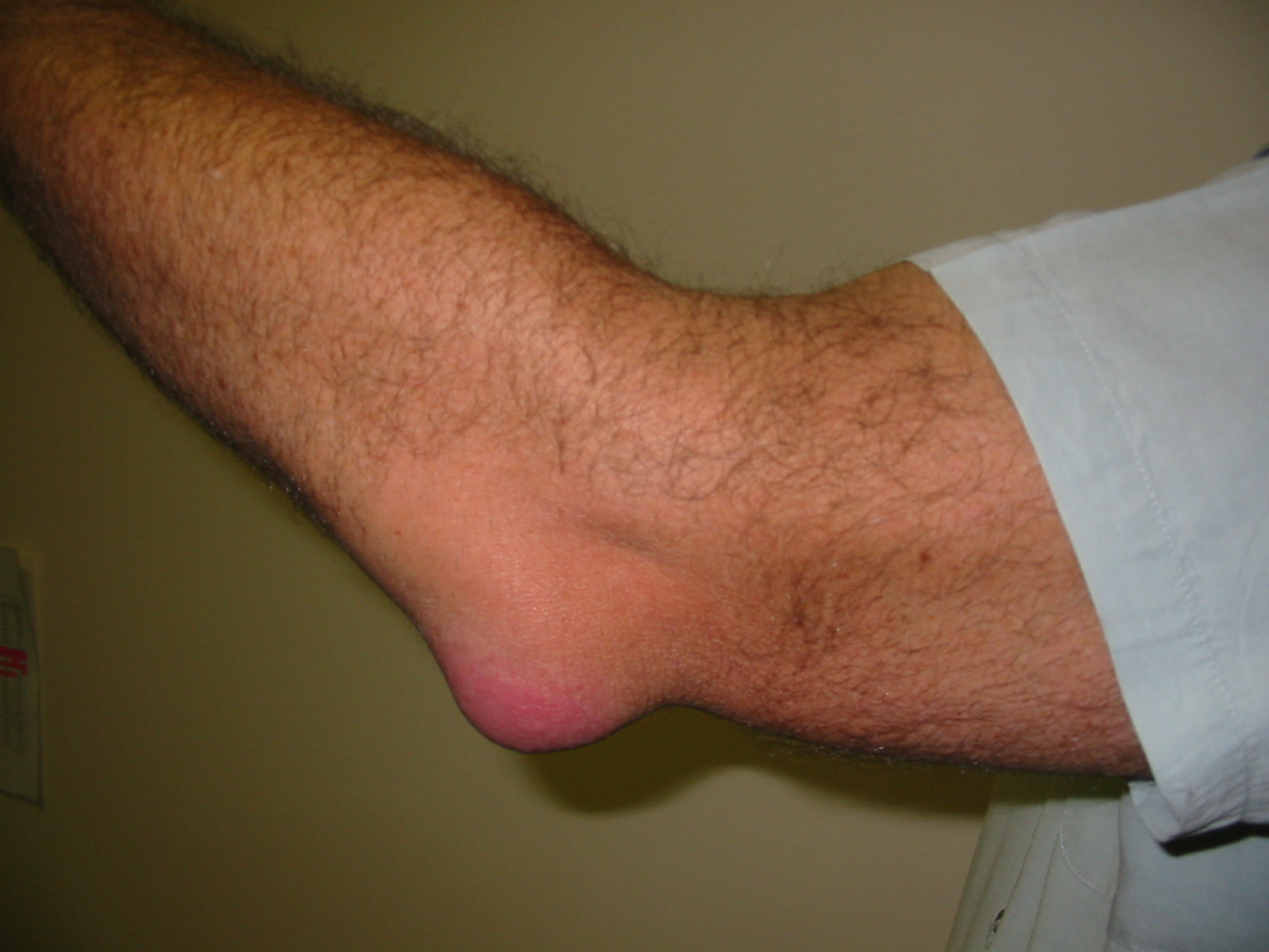
- Focal swelling on Posterior Aspect of Olecranon → Suggests Olecranon Bursitis
Olecranon Bursitis2
- Erythema
- Atrophy: Inspect the Biceps, Triceps, Brachioradialis, Wrist Flexors (attached to medial epicondyle) and Wrist Extensors (attached to lateral epicondyle)
- Deformities:
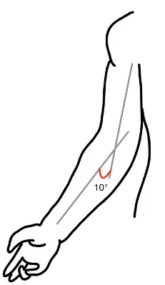
- Carrying Angle (Normal 5-10° in men, 10-20° in women): Angle of forearm with humerus when patient stands with arms at side, elbows extended, and palms facing forwards.
- Inability to fully extend the elbow → Suggests Flexion contractures
- Skin Changes or Scars:
- Rheumatoid Nodules: Rubbery nodules overlying olecranon
- Gouty Tophi: Hard nodules overlying olecranon
Palpation
AFMC level Pre-clerkship 3 · Clerkship 4
- Commenting on: TESTCA – Tenderness, Effusions, Swelling, Temperature, Crepitus, & Atrophy
- Palpate Posteriorly:
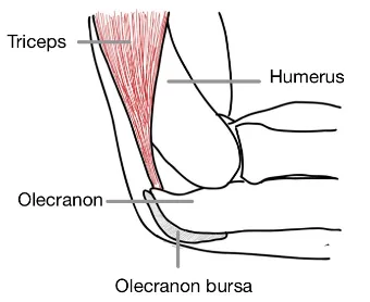
- Triceps
- Olecranon process
- Olecranon bursa
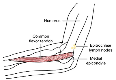
- Palpate Medially:
- Medial triceps border
- Epitrochlear Lymph Nodes (above medial epicondyle)
- Medial epicondyle
- Common flexor tendon (attached to medial epicondyle)
- Tenderness at medial epicondyle & common flexor tendon → Medial epicondylitis (Golfer’s Elbow)
- Palpate Anteriorly:
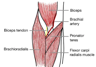
- Bicep
- Biceps Tendon (in antecubital fossa)
- Brachial pulse (medial to biceps tendon)
- Palpate Laterally:
- Lateral epicondyle
- Palpate for Elbow Effusion: Palpate lateral para-olecranon for effusion between lateral epicondyle and olecranon
- Common extensor tendon (attached to lateral epicondyle)
- Tenderness at lateral epicondyle & common extensor tendons → Lateral epicondylitis (Tennis Elbow)
- Radial head (should slip under thumb with elbow flexion to 90° and clinician’s thumb just distal to lateral epicondyle, with pronation and supination of forearm)
- Pediatrics: Pulled Elbow Syndrome – Radial Head Subluxation: Radial head is slipped out of normal position. Common in children under 5 years old because of relative strength of annular ligament.
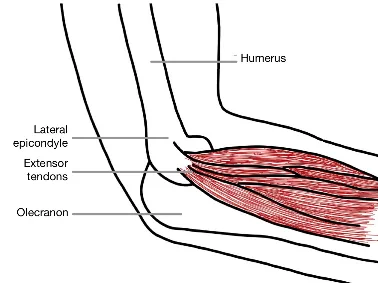
Why Check: Palpation can be very helpful to determine source of pain. Palpate for tenderness and swelling in a systematic manner, to assess for potential fractures and other abnormalities. Gout can present in the elbow as well, albeit more rarely.
Range of Motion (ROM)
AFMC level Pre-clerkship 3 · Clerkship 4
- Approach: Perform Active ROM, and then Passive ROM if Active ROM is limited. These can be integrated with examiner guiding the elbow beyond patient’s active ROM.
- Screening Test – Crepitus: With hand over elbow, move elbow joint passively
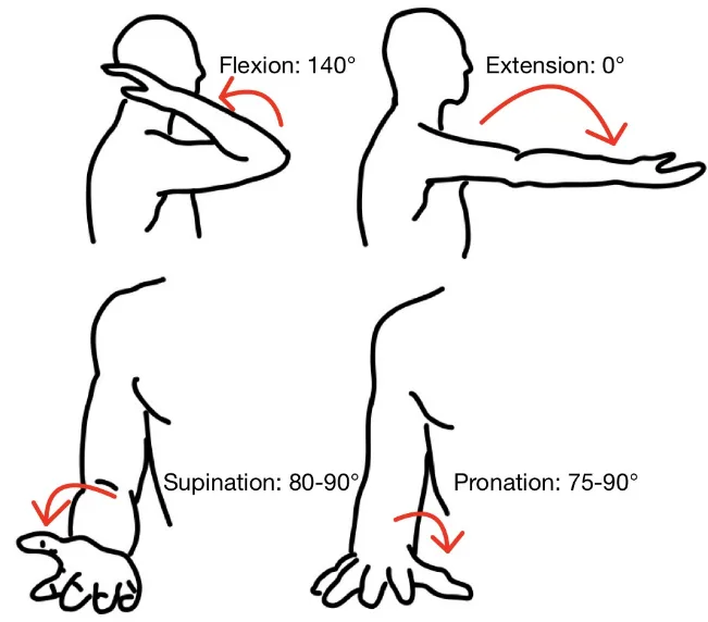
- Flexion (140° ± 10°)
- Extension (Full extension 0°, up to 10° hyperextension normal esp. in women) → inability to fully extend elbow is abnormal, may suggest elbow joint effusion or prior trauma (e.g. fracture)
- Pronation (75-90°) & Supination (80-90°): With arms at sides, elbows flexed and thumbs up, turn palms down (pronation), and then turn palms up (supination)
Power Assessment
AFMC level Pre-clerkship 3 · Clerkship 4
- Elbow Flexion with Forearm Supination: Patient makes a boxer pose; brace shoulder, grasp the forearm and apply an extensor force; patient resists motion.
- Elbow Extension with Forearm Supination: Patient makes a boxer pose; brace shoulder, grasp the forearm and apply a flexor force; patient resists motion.
- Forearm Pronation with Elbow Flexed to 90 degrees: Elbow flexed to 90°, palms up. Brace forearm with wrist in neutral position and apply supination force; patient resists motion.
- Forearm Supination with Elbow Flexed to 90 degrees, Arms at Side: Elbow flexed to 90°, palms down. Brace forearm with wrist in neutral position and apply pronation force; patient resists motion.
- Reporting: For each muscle group tested, assess per the Medical Research Council Power Grading. If it is all 5/5, you can say “Power was 5/5 bilaterally in both upper and lower limbs”, and report specific deficits. NOTE: This scale is detailed in Chapter 31 – Screening Neurological Examination
Special Tests:
Elbow Stability
Antero-posterior stability – [1, 3]
Say: I will assess the elbow for signs of instability.
Do: Support and stabilize elbow flexed at 30°. Grasp forearm with other hand.
- MCL/UCL Stability: Apply a valgus (outward) force against forearm. Assess for excess laxity with valgus force.
- LCL/RCL Stability: Apply a varus (inward) force against forearm. Assess for excess laxity with varus force.
- Anteroposterior Stability: Now flex elbow to 90 degrees. While stabilizing forearm with one hand, push and pull humerus with other hand. There should be no movement.
Report: No excess laxity with either a valgus or varus force applied – no signs of MCL or LCL instability.
Why Check: Excess laxity with a valgus force indicates a Medial (Ulnar) Collateral Ligament injury, while excess laxity with a varus force indicates a Lateral (Radial) Collateral Ligament injury. Movement anteriorly or posteriorly of humerus with elbow flexed to 90 degrees indicates antero-posterior instability.
Lateral Epicondylitis (Tennis Elbow) Test
Say: I will assess for signs of tennis elbow. Please let me know if you feel any pain, and if so, where.
Do: Palpate for tenderness at lateral epicondyle and along the common extensor tendon
- Extend patient’s elbow with wrist extended/cocked backwards.
- Apply a force at the wrist while the patient resists to maintain wrist extension. Look for signs of pain at lateral epicondyle or common extensor tendon.
- Patient then relaxes wrist and keeps elbow extended. Examiner pushes wrist into flexion to stretch the common extensor tendon, without patient resistance. Look for signs of pain at lateral epicondyle or common extensor tendon.
Report: No tenderness at lateral epicondyle or common extensor tendon – lateral epicondylitis test is negative.
Why Check: Tenderness at lateral epicondyle, tenderness at common extensor tendon, or pain at lateral epicondyle with active resistance or passive wrist flexion is suggestive of tennis elbow (lateral epicondylitis).
Medial Epicondylitis (Golfer’s Elbow) Test
Say: I will assess for signs of golfer’s elbow. Please let me know if you feel any pain, and if so, where.
Do: Palpate for tenderness at medial epicondyle. Elbow extended with wrist flexion.
- Extend patient’s elbow with wrist flexed (fingers pointing down)
- Apply a force at the wrist while the patient resists to maintain wrist flexion. Look for signs of pain at the medial epicondyle or common flexor tendon.
- Patient relaxes wrist and keeps elbow extended. Examiner pushes wrist into extension to stretch common flexor tendon, without resistance. Look for signs of pain at medial epicondyle or common flexor tendon.
Report: No tenderness at medial epicondyle or common flexor tendon – medial epicondylitis test is negative.
Why Check: Tenderness at medial epicondyle, tenderness at common flexor tendon, or pain at medial epicondyle with active resistance of wrist extension is suggestive of golfer’s elbow (medial epicondylitis). The Medial (ulnar) Collateral ligament is strongly attached to Medial Epicondyle, where an excess flexion injury or posterior elbow dislocation can cause avulsion fracture of the medial epicondyle.
Tinel’s Sign
AFMC level Pre-clerkship 3 · Clerkship 4
Say: I will examine the Cubital Tunnel in the elbow by gently tapping on an area on the inside of the elbow for 30 seconds. Let me know if you feel any pins and needles during the test, and if so, where.
Do: With patient elbow flexed at 90°, tap over ulnar nerve in the groove between the olecranon process and medial epicondyle for 30 seconds. Look for pain or paresthesia (pins and needles) radiating to 4th/5th fingers.
Report: No pain or paresthesias over ulnar nerve sensory distribution in hand – Tinel’s Sign negative.
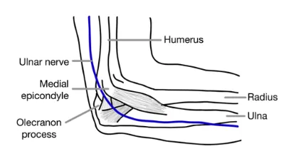
Why Check: Pain or paresthesias over ulnar nerve sensory distribution in hand (4th/5th fingers) is indicative of Cubital Tunnel Syndrome – caused by irritation or entrapment of the ulnar nerve in the cubital tunnel (groove between medial epicondyle and olecranon).
Part 3: Select Special Tests – Not Part of Routine Elbow Exam But Seen in Practice:
Elbow Flexion Test
Say: I will continue to examine the Cubital Tunnel in the elbow with movement of the arms. Please let me know if you feel any pins and needles during the test, and if so, where.
Do: Patient maximally flexes elbow for 60 seconds with wrists extended (to avoid compressing median nerve at wrist which can confound a positive test with Carpal Tunnel Syndrome). Look for paresthesias radiating to 4th/5th fingers.
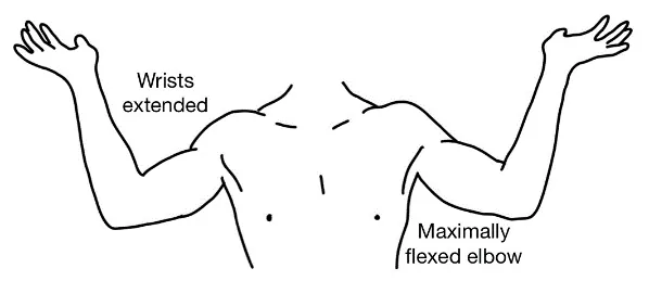
Report: No paresthesias developed over ulnar nerve distribution in hand – Elbow Flexion Test negative.
Why Check: Paresthesias over ulnar nerve sensory distribution in hand (4th/5th fingers) is indicative of Cubital Tunnel Syndrome – caused by irritation or entrapment of the ulnar nerve in the cubital tunnel (groove between medial epicondyle and olecranon).
Helpful Videos to Review:
- Video 1: Elbow Exam – McMaster MSK Examination Series (Link: ELBOW EXAMINATION - McMASTER UNIVERSITY)
- Video 2: Stethopedia Elbow Examination Video (Link: https://videopress.com/v/TDuYi3mo)
- Video 3 and 4: Bates’ Visual Guide to the Physical Exam – Anatomy Review and Examining the Elbows (Link: https://www.medportal.ca/ug/educational-resources/bates/lists/bates/musculoskeletal)
References
-
Coombes BK, et al. Effect of corticosteroid injection, physiotherapy, or both on clinical outcomes in patients with unilateral epicondylalgia: a randomized controlled trial. JAMA. 2013;309(5):461-469. ↩
-
Bursitis_Elbow_WC.JPG. Wikimedia Commons. Accessed May 2, 2023. https://upload.wikimedia.org/wikipedia/commons/6/6e/Bursitis_Elbow_WC.JPG ↩