Integration Foundation
Chapter 40Ophthalmology: Essential Anatomy and Clinical Skills
Part 1: Ophthalmology – Key Anatomical Principles and Terminology
Extraocular Muscle Organization
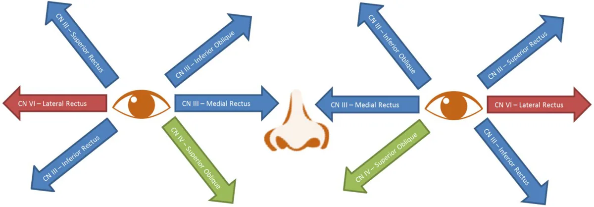
- There are 6 extraocular muscles in each eye, which form the rotator cuff of the eye. The eye performs 6 movements primarily, with extraocular muscle organization and innervation as detailed in the table below:
| Movement | Muscles | Innervation |
|---|---|---|
| Adduction (inward) | Medial Rectus | CN III |
| Abduction (outward) | Lateral Rectus | CN VI |
| Supraduction (upward) | Superior Rectus + Inferior Oblique | CN III |
| Infraduction (downward) | Inferior Rectus + Superior Oblique | CN III + CN IV |
| Intorsion (rotate toward nose) | Superior Oblique | CN IV |
| Extorsion (rotate away from nose) | Inferior Oblique | CN III |
- Note: The oblique muscles’ primary action is rotating the eye. Inferior Oblique (primarily extorsion, as well as supraduction) and Superior Oblique (primarily intorsion, as well as infraduction) correct the outward/inward motions of the superior and inferior rectus.
- Superior and Inferior Obliques perform corrective actions as the head rotates to keep eyes gyroscopically stable
- As a rotator cuff, extraocular movements are a combination of subtle movements of all muscles comprising the cuff.
Direction of Action of the Extraocular Muscles (CN III, IV, VI)
- Extraocular muscle innervation memory aid: (LR₆SO₄)³ → Lateral Rectus = CN VI, Superior Oblique = CN IV; others innervated by CN III.
- Direction of movement:
- What Cranial Nerves control the opening and closing of the eyelids?
| Movement | Muscle | Innervation |
|---|---|---|
| Opening Eyes | Levator Palpebrae Superioris | CN III (Oculomotor) Eyelid opening also controlled by Sympathetic nervous system – i.e. a Horner’s Syndrome gives a mild Ptosis |
| Closing Eyes | Orbicularis Oculi | CN VII (Facial – Temporal and Lacrimal branches) |
- What is hardest to detect clinically? Lesions involving superior and inferior oblique muscles as these rotate the eye.
- Superior Oblique: Lesions occur with Cranial Nerve IV and present with history of diplopia when looking down.
- Inferior Oblique: Lesions occur with Facial Fractures and present with history of diplopia when looking up.
Pupillary Light Reflex and Pupillary Accommodation Reflexes
- Pupil size is a balance between Sympathetic dilation of pupil and Parasympathetic constriction of pupil.
Parasympathetic Response
- Purpose: When light enters one pupil, the same pupil (direct response) and the contralateral pupil (consensual response) will both constrict to ↓ light entering both eyes.
- Mechanism:
- Step 1: Afferent pathways begin with the optic nerve (CN II), with nasal fibres crossing at the optic chiasm
- Step 2: Some fibres travel through the Lateral Geniculate Nucleus (LGN, Thalamus) and sends signal to the visual cortex
- Step 3: Some fibres travel to the pretectal area of the midbrain, which sends signals to the Edinger-Westphal Nuclei. Both the ipsilateral AND contralateral nuclei will constrict the eyes to cause direct and consensual response.
- Step 4: The parasympathetic fibres travel as the outer portion of the oculomotor nerve (CN III)→ ciliary ganglion → short ciliary nerves → pupillary sphincter muscles → constriction of pupils
Key Clinical Correlations:
- Relative Afferent Pupillary Defect (RAPD – Marcus Gunn Pupil): In swinging flashlight test, shining light in affected eye causes ↓ pupillary constriction in both eyes; shining light in unaffected eye will cause normal pupillary constriction in both eyes. A dilation of one pupil during swinging flashlight test suggest a positive RAPD, which corresponds to a retina or optic nerve injury. This is extremely important, as it is often only objective finding in optic neuritis or a tumor compressing on optic nerve on exam.
- Anisocoria: Unequal pupil size. Can be caused by 3 categories of pathologies.
| Pathology | Clinical Finding |
|---|---|
| Parasympathetic issues | Greater anisocoria in bright light (Parasympathetic Nervous System responsible for constricting the pupil). |
| Sympathetic issues | Greater anisocoria in dim light (Sympathetic Nervous System responsible for dilating the pupil). |
| Physiologic anisocoria | Found in ~10-20% of population, benign. Will have equal anisocoria in dim and bright light. |
- A differential for anisocoria greater in the light includes:
- Adie’s Tonic Pupil: A lesion of efferent parasympathetic innervation on one side → Anisocoria (unequal pupillary size) with tonically dilated pupil only slowly reactive to light; for the unaffected eye, observe normal reactivity.
- Oculomotor Nerve Palsy (CN III): Due to CN III’s 3 functions (eye movement, opening of eyelid, pupil constriction), CN III palsy will lead to a “down and out” affected eye, ptosis, and anisocoria (unequal pupil size). As parasympathetic fibres form outer portion of CN III, aneurysms or tumours compressing CN III will lead to a CN III palsy involving the pupil (pupil dilated when light shone) versus ischemia involving CN III will lead to palsy sparing the pupil (pupil is reactive to light).
- Hutchinson’s Pupil: Partial Oculomotor Nerve palsy → deficits to parasympathetic response without deficits in extraocular muscles due to parasympathetic fibers running through oculomotor nerve periphery; can be seen before full CN III palsy.
- Cycloplegic Eye Drops: e.g. Tropicamide – short acting anticholinergic medication → paralysis of pupillary sphincter muscles, allowing sympathetic tone to dilate pupil in affected eye as a purposeful Anisocoria to facilitate examination.
Sympathetic Response
- Purpose: Acting to dilate the pupil and ↑ light entering both eyes
- Mechanism:
- Step 1: Hypothalamus sends signals down to C8/T1 spinal level
- Step 2: Superior cervical ganglion travels alongside the Internal Carotid Artery to the eye
- Step 3: Innervation to Iris dilator muscle and Muller’s Muscle (additional 2mm elevation of the superior eyelid)
Key Clinical Correlations:
- Horner’s Syndrome: Ipsilateral oculosympathetic pathway lesion
- Horner’s Syndrome Triad: Miosis (of affected side), partial ptosis (Muller’s Muscle lesion), facial anhidrosis
- Causes: Idiopathic; brainstem stroke; carotid artery dissection; neoplasm (mass effect e.g. Pancoast tumor)
Pupillary Accommodation Reflex
- Purpose: To adjust refractive index of eye to focus image, whether object being focused on is near or far away.
- Mechanism: Same apparatus as the Parasympathetic pathway, with the addition of ocular motor control neurons from visual association cortex which sends corrective signals to control the CILIARY MUSCLES – changing SHAPE of the lens.
- Nearby Object: Signals to contract the ciliary muscles and make the lens more curved with the focal length shortened.
- Distant Object: Signals to relax the ciliary muscles and make the lens less curved with the focal length increased.
Key Clinical Correlation:
- What is the interplay between Pupillary Accommodation, Pupillary Light Response, and Exam Setting Lighting?
- When Testing Pupillary Light Reflex: Ask patient to focus on a target in distance during swinging flashlight test –prevents pupillary constriction due to accommodation for nearby objects interfering with the test.
- When Testing the Pupillary Accommodation Reflex: Use moderate ambient lighting to prevent pupillary constriction due to the ambient light interfering with the test.
Visual Field Testing
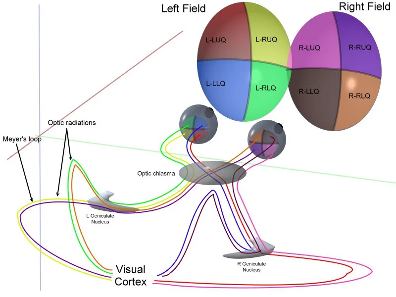
Key Concepts of Visual Fields (see right)
- Temporal visual field falls on nasal part of retina; nasal visual field falls on temporal part of retina.
- Macula most sensitive retinal area (fovea at centre of macula) and is disproportionately over-represented in the visual field.
- Blind-spot is at optic disc due to absence of photoreceptors at this site where optic nerve exits. The visual cortex will later “fill this in”.
- Nasal fibres of optic nerve cross at optic chiasm i.e. temporal visual fields.
- At Lateral Geniculate Nucleus (LGN, Thalamus), left visual field unified at right LGN, right visual field unified at left LGN.
- Visual information split into quadrants via optic radiations, with upper quadrants represented by Meyer’s Loops.
- Optic radiations travel to Primary Visual Cortex, where information integrated to reproduce visual field, in broad terms by parallel processing and a topographical mapping arrangement conserved throughout visual sensory system.
Other Common Ophthalmologic Lesions – Intraocular Defects Affecting Visual Field:
- Altitudinal Defect: Entire top or bottom half of field involved; more likely to be an ocular disease vs cortical
- Arcuate Scotoma: Arcs from the blind spot around fixation to the nasal side – a glaucomatous defect
- Central Scotoma: Defects in the retina (the closer the lesion to the macula, the larger the scotoma)
Key Abbreviations Used in Ophthalmology Clinical Practice
| Type | Abbrev | Meaning |
|---|---|---|
| Anatomic | RE | Right Eye |
| OD | Oculus Dexter (Right Eye in Latin) | |
| LE | Left Eye | |
| OS | Oculus Sinister (Left Eye in Latin) | |
| BE | Both Eyes | |
| OU | Oculus Uterque (Both Eyes in Latin) | |
| Vision | VA | Visual Acuity |
| CF | Counting Fingers | |
| HM | Hand Motions | |
| LP | Light Perception | |
| NLP | No Light Perception | |
| PH | Pinhole | |
| Diagnoses | ET | Esotropia (Eye turned nasally) |
| XT | Exotropia (Eye turned temporally) | |
| LHyperT / RHyperT | L or R Hypertropia (Eye turned upwards) | |
| LHypoT/ RHypoT | L or R Hypotropia (Eye turned downwards) | |
| ACG | Angle Closure Glaucoma | |
| AMD | Age-Related Macular Degeneration | |
| PVD | Posterior Vitreous Detachment | |
| NVD | Neovascularization of the disc | |
| NVE | Neovascularization Elsewhere (of fundus) | |
| NVI | Neovascularization of the Iris | |
| Exam | VF | Visual Field |
| SLE | Slit Lamp Examination | |
| IOP | Intraocular Pressure | |
| V cc | Visual acuity with glasses | |
| V sc | Visual acuity without glasses |
Part 2: Focused Ophthalmological History
HPI, Assoc. Symptoms
Overview Questions
(explore different sections below if positive)
- What do you see? Any blurry vision, vision loss, visual field loss, double vision, flashes or floaters
- How do your eyes feel? Any pain, itchiness, photophobia
- How do your eyes look? Any redness, bulging
- Any discharge? Any purulence, clear discharge, involving one or both eyes
- Any other symptoms? Any headache, scalp tenderness, jaw claudication, tinnitus, motor or sensory deficits?
Discharge
- Conjunctivitis (Pink Eye) → Can be bacterial (unilateral purulent discharge), viral (unilateral watery discharge), or allergic (chronic bilateral itching and tearing).
- Wear gloves, and after examination wash with soap and wipe down exam area including the chair as viral is conjunctivitis is very contagious
Blurry Vision
- Dry eyes → Worse when reading/watching TV, near end of day
- Cataracts → Generalized blurriness, glaring at night
- Glaucoma → Tunnel vision (peripheral vision impacted)
- AMD (Age-Related Macular Degeneration) → Central distortions, central scotoma (partial loss)
- Diabetic retinopathy → Blurred central vision
Diplopia (Double Vision)
- Monocular diplopia (persists when unaffected eye is covered): intraocular pathology (e.g. cataracts)
- Binocular diplopia (disappears when either eye is covered): neural or extraocular muscle pathology (e.g. cranial nerve palsy)
Painful or Non-Painful
- Painful vision changes: Infection, inflammation (e.g. corneal infection, uveitis, acute angle closure glaucoma)
- Painless vision changes: Cataract, Age-Related Macular Degeneration (AMD), chronic glaucoma
Flashes & Floaters
- Posterior Vitreous Detachment – vitreous humor losing gelatinous property over time, tugs on retina leading to flashes and floaters. These can progress to retinal tears and detachments.
Vision Loss
- Key Question: Does it feel like there is a curtain coming down?
- Referring: If loss of visual fields → retinal detachment until proven otherwise; refer ASAP
Headache and Scalp Tenderness
- Giant cell arteritis: presents with headache, scalp tenderness, jaw claudication (difficulty chewing), constitutional symptoms. Refer ASAP as vision and life-threatening.
Contacts Use
- Type of contacts (daily, weekly, month change), length of wear per day (recommended <10 hours daily), method of cleaning, contact hygiene (bad if wearing to sleep or shower or pool) → poor hygiene increases risk of corneal infections from bacteria and fungi.
Past Medical History (PM Hx)
- Past ocular history: eye conditions (e.g. glaucoma), eye trauma
- Rheumatologic Diseases: Can cause iritis (uveitis) – ask about joint pain, psoriasis, etc.
- Scleroderma (Progressive Systemic Sclerosis): Contributes to dry eyes
- Diabetes: Diabetic retinopathy, ask about duration of diabetes
- Hypertension: Hypertensive retinopathy, also risk factor for many eye conditions
- Thyroid Conditions: Graves’ Disease (Hyperthyroidism Autoimmune) causes Exophthalmos (bulging of eye), restrictions in extraocular movements, which cause diplopia
Past Surgical History (PS Hx)
- Strabismus Surgery: As a child, patient might remember having a “lazy eye” that was surgically corrected
- Laser Vision Correction, Cataract Surgery
Family History (FM Hx)
- Blindness; Conditions such as retinal detachment; Glaucoma; AMD (Age-Related Macular Degeneration)
Social Hx
Occupation Hx
Heavy industry – patients may present with foreign body in the eye (e.g. metal). Ask if they grind metal.
Medications Hx & Allergies
Medications
- Glaucoma: Ask – are you on any eye drops?
Allergies
- Seasonal Allergies: Can cause allergic conjunctivitis
Part 3: Ophthalmic Examination
Visual Acuity (VA) Measurement (Adults and Children ≥5 years old)
AFMC level Pre-clerkship 3 · Clerkship 4
Say: I will assess your vision reading an eye chart. If you have glasses or contacts, please keep them on as I need to test your corrected vision. Please squint if you need to. Please cover one eye.
METHOD 1 – POCKET SNELLEN CHART:
Setup: Holding pocket Snellen chart to patient’s nose 16 inches from patient’s nose (slightly more than a card length) to ensure an appropriate reading distance.
Do: With one eye covered, read from bottom of card up, reading the lowest line they can. Then, cover the other eye, and read from top down, until they reach the lowest line they can (this prevents the patient from remembering the lines which would obscure results).
Report: The patient has a vision of 20/20 in both the left and right eyes.
METHOD 2 – PROJECTOR SNELLEN CHART:
Setup: Have patient 20 feet (6 meters) away from chart.
Do: With one eye covered, read lowest line they can. Then cover the other eye and read the lowest line (in reverse order).
Report: The patient has a vision of 20/20 in both the left and right eyes.
CONDITIONAL TEST – IF PATIENT CANNOT SEE THE 20 / 400 (big E) LINE:
Say: I will now ask you to count fingers at a distance, and a couple other vision tests if needed.
Do – Counting Fingers (CF): Hold up your fingers at varying distances (e.g. 5 metres, 4 metres, etc.) and ask the patient to count fingers. Record the maximum distance they can count fingers between 1 and 5 metres (e.g. VA = CF 5 metres or VA = CF 1 metre)
Do – Hand Motion (HM): If patient unable to count fingers at 1 metre, wave your hand at 1 metre away and ask if the patient can see this. Record if the patient can see Hand Motion (e.g. VA = HM)
Do – Light On/Off Light Perception (LP or NLP): If patient unable to Count Fingers or see Hand Motion at 1 metre away, shine a light in eye and check if the patient can see the light. Record if the patient can see the light (VA = LP) or are unable to see the light (VA = NLP).
Report: The patient is unable to count fingers but is able to see Hand Motion.
Documentation: (e.g. VA cc OD 20 / 40, OS CF at 2 feet)
- Document on chart cc for corrected vision (with glasses or contacts), sc for uncorrected vision.
- If pinhole available, document if vision improves with pinhole.
- What does 20 / 100 – 2 mean? A patient sees at 20 feet what a normal sighted patient sees at 100 feet; with 2 letters incorrect on that line.
- What does 20 / 100 + 2 mean? A patient sees at 20 feet what a normal sighted patient sees at 100 feet; with 2 correct and 3 incorrect on the line below.
Why Check: Visual Acuity is one of the 3 “vital signs” in ophthalmology, along with pupillary response to light and intraocular pressure. Visual acuity is a test of macular function and optic nerve function.
- Design of Snellen Charts: 20/20 - Numerator is test distance; denominator is distance that a normal sighted person able to identify letter → e.g. 20/70, patient can discriminate letter from 20 feet, whereas a normal person can do so at 70 feet. Can use pocket Snellen chart in limited exam settings.
- Ametropia: Refractive error in vision
- Myopia: Near sightedness – sharp image formed in front of retina; refractive power too strong for axial length of eye
- Hyperopia: Far sightedness – sharp image formed behind retina; refractive power not strong enough for axial length of eye
- Why does Squinting improve visual acuity? Squinting ↓ light rays entering eye → ↓overall blurring of image. If squinting improves vision, ↑ suspicion refractive error is contributing to ↓ visual acuity. If squinting does not improve vision, ↑ suspicion of organic problem in visual pathway contributing to the ↓ acuity. This is why all abnormal visual acuity is tested with pinhole (PH) to eliminate any refractive error contributing to low visual acuity.
- What is legal blindness? 20 / 200 best corrected visual acuity (wearing distance glasses) or worse in better eye.
- What is legal requirement of vision to hold Ontario G or M license? 20 / 50 best corrected visual acuity in their better eye.
- Note: flight license 20 / 30 best corrected visual acuity or better in better eye and uncorrected is not less than 20 / 200.
- Pediatrics: For children 3-5 years old – use an Allen Picture Chart; for children 0-3 years old – assess ability to Fix and Follow a toy – described in Pediatric Ophthalmology section (below)
Pupillary Response to Light
AFMC level Pre-clerkship 3 · Clerkship 4
Say: I will assess each eye’s response to light. Please remove your glasses and keep both eyes open during this test.
Do: Inspect pupil size (Snellen Pocket card tends to have a guide). ≤1 mm difference between two eyes is normal. Using a flashlight, shine light in one eye. Look for pupillary constriction both direct and consensual (part of the Pupillary Light Reflex). Repeat in other eye. Then, swing flashlight back and forth between both eyes, each pupil should constrict (no relative afferent pupillary defect).
Report: Pupils are equal and reactive to light, there is no relative afferent pupillary defect.
Why Check: Pupillary Response to Light is one of the 3 “vital signs” in ophthalmology, along with Visual Acuity and Intraocular Pressure.
- Pupillary Response: PERRLA (Pupils Equal Round and Reactive to Light & Accommodation) test has high sensitivity for optic nerve disease, ipsilateral brain herniation, and Posterior Communicating Artery (Circle of Willis) aneurysm. It also has high sensitivity & specificity for Horner’s Syndrome.
- Positive relative afferent pupillary defect (RAPD): Dilation of one pupil during swinging flashlight test suggests a retina or optic nerve injury – extremely important as often the only objective finding on exam in optic neuritis or a tumor compressing on the optic nerve.
Intraocular Pressure (IOP) Measurement
Say: I will now freeze the eye with a local anesthetic, it will last 10 minutes. Please keep the eye open and look at one point in the distance. I will then measure the eye pressure by tapping gently, which is painless.
Setup: Use a Tonopen – commonly available in Emergency Departments
Do:
- Freezing: Freeze eye with 1 drop of local anesthetic (e.g. Alcaine) – lasts about 10 minutes.
- Calibrate Tonopen: Point Tonopen towards floor, press button twice, initially “CAL” will appear on screen followed by “UP”, then point Tonopen towards sky.
- Approach: Do not push on eyelid (artificially elevates IOP), hold Tonopen like a pen, brace hand on cheek for stability, tap lightly on centre of cornea multiple times (each time will “beep”) until a number appears on screen.
Report: The Intraocular pressure measurement is 15 mmHg, which is normal.
Why Check: Intraocular Pressure is one of the 3 “vital signs” in ophthalmology, along with Visual Acuity and Pupillary Response to Light. Intraocular pressure is a balance between the rate of production of aqueous humor of the eye, resistance to aqueous outflow through trabecular meshwork and Schlemm’s Canal, and Episcleral venous pressure.
- Normal: 10-21 mmHg; if a blockage occurs, Intraocular pressure (IOP) can quickly rise to 60-70 mmHg e.g. in acute angle closure glaucoma – an ophthalmologic emergency (may also present with headache, halos around lights, severe eye pain and nausea/vomiting)
External Ocular Exam
(See Ch 7 – Head and Neck Exam)
Say: I will inspect each eye quickly, before we proceed to the other steps of the exam.
Setup: Look at the eye without any tools in ambient lighting.
Do:
- Shape of Eye/Eyelid: Check for Ptosis (drooping of eye), Exophthalmos (proptosis – protrusion of the eye), Entropion (eyelids folding inwards, eyelashes irritate cornea – Surgically corrected) or Ectropion (eyelid turned outwards – Surgically corrected).
- Redness: Check for Conjunctivitis (generalized red vessels on conjunctiva); Iritis or Acute Angle Closure Glaucoma will have ciliary flush (red vessels just around the cornea)
- Specific Lesions: Check for Chalazion (cyst of the eyelid), Papilloma (benign epithelial tumor)
- Other Structures: Inspect the Lacrimal Gland, Puncta, Canaliculi, Nasolacrimal Sac and Duct
Report: There are no abnormalities in the external structures of the eye.
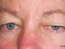
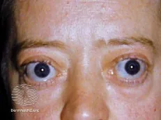
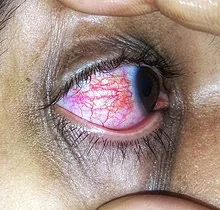
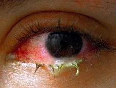
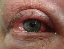
Ptosis1, Exophthalmos2, Viral Conjunctivitis3, Bacterial Conjunctivitis4, Allergic Conjunctivitis5
Why Check: Common part of the ophthalmic examination alongside the 3 “vital signs” of ophthalmology.
- Globe Rupture = Ophthalmologic Emergency (the STEMI of Ophthalmological Practice): A communication between the outside world and the inside of the eye. This is a can’t miss diagnosis where the external ocular exam is an important part. Signs include a Sunken eye, pupil irregularity, prolapse of intraocular contents outside the eye, bullous subconjunctival hemorrhage, and hyphema (collection of blood in anterior chamber of the eye). If you suspect a Globe Rupture:
- Document agent of injury, when injury occurred, and tetanus status.
- Once diagnosis made, 1) Check vision → 2) patch eye with shield → 3) refer to ophthalmology for surgery immediately. Do NOT check Intraocular Pressure or do further exams.
- Ptosis: Consider Cranial Nerve injury or sympathetic nerve injury (e.g. Horner Syndrome with triad of Miosis of affected side, partial ptosis due to Muller’s Muscle lesion, and facial anhidrosis
- Exophthalmos: Sign of Thyroid disease (Ch 15 – Thyroid Exam – MF3)
Acute conjunctivitis can represent:
- Viral conjunctivitis: Classically appears as a pink or red colour, with minimal-to-no itching and watery discharge, which usually begins unilateral but may progress to bilateral. Ask about sick contacts, where contact with another person with red-eye mildly increases the likelihood of viral conjunctivitis (LR+ 2.5, LR- 0.89). Ask if eyelids are stuck together on awakening which mildly decreases the likelihood of viral conjunctivitis (LR+ 0.72, LR- 3.5). On exam, look for mucopurulent ocular discharge (LR+ 0.47, LR- 2.7) which moderately decreases the likelihood of viral conjunctivitis. Also look for pharyngitis (LR+ 5.4-9.9, LR- 0.48-0.48) and preauricular lymphadenopathy (LR+ 2.5-5.6, LR- 0.73-0.89) which moderately increase the likelihood of viral conjunctivitis.
- Bacterial conjunctivitis: Classically appears as a pink or red colour, with minimal-to-no itching and mucopurulent discharge which is usually unilateral. Ask about sick contacts, where contact with another person with red-eye moderately reduces the likelihood of bacterial conjunctivitis (LR+ 0.40). Ask if eyelids are stuck together on awakening (LR+ 1.4, LR- 0.29) which mildly increases the likelihood of bacterial conjunctivitis. On exam, assess for ocular discharge, where watery discharge greatly reduces the likelihood of bacterial conjunctivitis (LR+ 0.03), as well as mucopurulent discharge (LR+ 2.1, LR- 0.37) and otitis media (LR+ 2.5, LR -0.84) which mildly increase the likelihood of bacterial conjunctivitis. look for pharyngitis (LR+ 0.10-0.19, LR- 2.1-2.14) and preauricular lymphadenopathy (LR+ 0.18-0.40, LR- 1.1-1.4) which moderately decrease the likelihood of bacterial conjunctivitis.
- Allergic conjunctivitis: Pink; Prominent itching; Watery discharge; bilateral. In allergic rhinitis, can also see infraorbital edema/darkening due to subcutaneous venodilation re. “allergic shiners”.
Confrontational Visual Fields
AFMC level Pre-clerkship 3 · Clerkship 4
Say: I will assess your vision in 4 quadrants in each eye by counting fingers. If you have glasses or contacts, please keep them on. Please stare at my nose during this test.
Setup: Sit at same level as patient. Key idea –you are the standard the patient is compared to i.e. symmetry should exist between you and patient to test the same visual field. When holding up fingers, use 0, 1 or 2 fingers in a plane perpendicular to axis formed by examiner and patient’s noses.
Do: The patient should close and cover one eye while you close opposite eye, so you have same field of vision. Hold out your hand in one quadrant equidistant between yourself and pt (fingers in same visual field). How many fingers am I holding up? Repeat in all 4 quadrants and for the other eye.
Report: The patient has a normal field of vision bilaterally in all 4 quadrants. If any visual field defects, record your findings using circle diagrams noting defects in quadrants of each eye.
Why Check: Alongside the 3 “vital signs” of ophthalmology – Visual Acuity, Intraocular Pressure and Pupillary Response to Light – Confrontational Visual Fields and Extraocular Muscle Movements are fundamental tests performed on each patient and part of the screening neurological examination (See Chapter 31 – Screening Neurologic Exam – MF4). By carefully testing the quadrants of the visual field of each eye, and drawing a diagram representing your findings as to where the deficit is, you can localize where the lesion is, as seen below:
| Where is the Lesion? | Visual Field Areas Lost | Visual Field Diagram | Visual Field Description |
|---|---|---|---|
| Partial lesion of left optic nerve (e.g. in Multiple Sclerosis) | Loss of central vision in the left visual field | 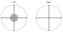 | Unilateral Central Scotoma in Left Visual Field |
| Complete section of left optic nerve | Loss of all vision in the left visual field | 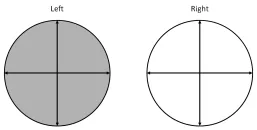 | Complete Blindness in Left Eye |
| Optic chiasm (commonly associated with pituitary adenomas) | Loss of temporal vision in both visual fields | 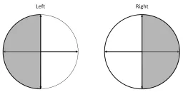 | Bitemporal Hemianopia |
| Left Optic Tract before Left Lateral Geniculate Nucleus | Loss of LUQ and LLQ in both visual fields | 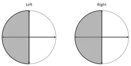 | Left Homonymous Hemianopia |
| Left Lower Optic Radiations | Loss of LLQ in both visual fields | 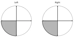 | Left Homonymous Lower Quadrantopia |
| Left Upper Optic Radiations (Meyer’s Loop) | Loss of LUQ in both visual fields | 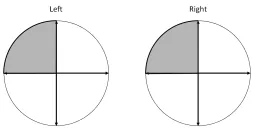 | Left Homonymous Upper Quadrantopia |
| Left Occipital Lobe | Loss of LUQ and LLQ in both visual fields, with macular sparing | 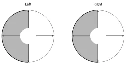 | Left Homonymous Hemianopia with Macular Sparing |
| Occipital contusion (often from backward falls) | Loss of central vision in both visual fields | 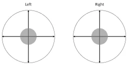 | Bilateral Central Scotomas |
Extraocular Muscle Movements
AFMC level Pre-clerkship 3 · Clerkship 4
Say: I will assess movements in each eye; please focus on my finger without turning your head.
Do: Move in a slow H pattern and observe eye movements of patient. As you move your finger slowly, observe a smooth pursuit where eyes have a conjugacy of gaze (align to focus on your finger). Give patient feedback if you notice them visibly moving their head as they track, as this does not allow you to test the more lateral aspects of their ocular movements. Ensure there are no restrictions in eye movements at extremes of gaze.
Report: I note conjugacy of gaze with smooth pursuit, no nystagmus or abnormal eye movements.
Why Check: Alongside the 3 “vital signs” of ophthalmology – Visual Acuity, Intraocular Pressure and Pupillary Response to Light – Confrontational Visual Fields and Extraocular Muscle Movements are fundamental tests performed on each patient and part of the screening neurological examination (See Chapter 31 – Screening Neurologic Exam – MF4). Specifically, testing the 6 cardinal extraocular movements also examines Cranial Nerves III, IV and VI (as detailed in screening neurologic examination).
Anterior Segment (Slit-Lamp) Exam
Say: I will inspect each eye more closely using a Slit Lamp. Would you please take a seat, and position the chin in the chin rest, and forehead against the top strap?
Setup: Room should have low ambient lighting. Ensure patient sitting upright comfortably with chin in chin rest, and forehead against forehead band. Adjust your own chair to comfortably perform exam.
Do:
- Key Idea: The Slit Lamp an autofocus such that light source and your lenses are at same focal point. In the slit lamp exam, you move focal point of device in space using controls to examine anterior structures; each slit lamp arranged slightly differently.
- To Start: Low-light and broad beam to begin, then narrow in. Examine one eye at a time.
- Exam Anterior→Posterior: Lid→ Lacrimal→ Lashes → Sclera → Conjunctiva → Cornea → Iris → Anterior Chamber → Lens
- Lids, Lashes and Adnexal Structures: Assess first to identify Blepharitis (flakes on lids), can flip upper lid for foreign bodies.
- Conjunctival and Scleral Examination: Ask patient to look left, right, up, down to view entire sclera. Identify any conjunctivitis or ciliary flush (red ring around cornea).
- Cornea: Assess for opacities or foreign bodies.
- Anterior Chamber: Assess for any hypopyon (white blood cells) or hyphema (red blood cells). Cells or Flare is seen in uveitis: best observed with short beam of high intensity light into centre of pupil at angle, at high magnification. Consider cells as specks of dust seen, flare as beam of projector.
- Lens: Assess for opacification suggesting cataracts
- Special Light Filters and Dyes – Cobalt Blue: Apply fluorescein (yellow dye) to the eye, blue light will show corneal epithelial defects suggesting a corneal abrasion (lights up green).
Report: No abnormalities of the anterior segment of the eye are seen on Slit Lamp Exam. No corneal abrasion seen after Fluorescein staining.
Why Check: The Slit-Lamp Exam allows for a focused exam of intraocular structures, alongside external examination and direct fundoscopy. Some common presentations to Emergency Departments where a Slit Lamp exam is helpful include:
1) Corneal Abrasion – A common presentation to Emergency Departments:
- What is it? Corneal epithelial defect from scratching the eye. Patient presents with red painful eye.
- Signs of corneal abrasion: Area of fluorescein uptake under blue light. Must rule out corneal ulcer which presents with corneal opacity.
- What should you do? Prescribe topical antibiotics (e.g. Moxifloxacin QID and Ciprofloxacin ointment nightly), Tylenol and Advil for pain. Avoid patching the eye, especially if patient wears contacts – contacts can be a growth medium for Pseudomonas, which favours anaerobic conditions.
Foreign Body – Visibly on eye surface: Common especially in industrial occupations, damaging glasses, etc.
- What should you do? Ensure tetanus is up to date. Must order an orbital X ray to rule out intraocular foreign body for every case of metal foreign body. To remove the foreign body, using topical anesthesia and a small gauge needle balancing your hand on the patient’s face, gently pry the foreign body off the cornea. Refer to ophthalmology if any rust ring remains.
Foreign Body –Under Upper eyelid: Common especially in industrial occupations, damaging glasses, etc.
- Signs of foreign body under the eyelid: Vertical corneal abrasions on cornea with fluorescein under blue light.
- What should you do?
- Flip Upper Eyelid: Have patient look down and resist blinking. Firmly grasp a cotton swab at cotton end as if holding a pen. Press swab against middle of eyelid firmly. This creates a fulcrum for eyelid to be flipped over. Grasp upper eyelashes → firmly move eyelid over the cotton swab fulcrum. Hold the eyelashes to keep in position.
- Remove foreign body: Using topical anesthesia and a small gauge needle, balance your hand on the face and gently pry the foreign body off cornea. Once finished, have patient look down and blink.
- Ensure tetanus is up to date. Must order an orbital X ray to rule out intraocular foreign body for every case of metal foreign body.
Direct Fundoscopy
AFMC level Pre-clerkship 2 · Clerkship 3
(Also explored in Ch 7 – Head and Neck Exam – MF1; Ch 14 – Diabetes Exam – MF3; and Ch 31 – Screening Neuro Exam – MF4)
Say: I will now inspect each eye using a direct ophthalmoscope. You will feel my hand touch the shoulder to keep the ophthalmoscope steady. Please look at an object on the wall in the distance at your eye level (preventing constriction due to pupillary accommodation reflex)
Setup: Room should still be dim. Stand facing the patient; if you’re examining the patient’s right eye first, use your right eye to look (vice versa). Adjust the scope’s focus (if you’re not wearing your nearsighted prescription glasses, may take more adjusting) by focusing on an object in room. Use low-moderate light intensity typically with medium aperture.
Do: Steady your hand on their shoulder. Shine the light from ophthalmoscope in patient’s eye; should see the red reflex. Follow reflex and slowly move toward patient. Find an artery and follow that towards the centre. You should come upon the optic disc (head of optic nerve, a confluence of retinal nerve fibers exiting the globe) and the optic cup (white centre of optic disc). Look for signs of optic disc swelling, and loss of venous pulsations. Follow the Superonasal Arcade, Inferonasal Arcade, Superotemporal Arcade, and Inferotemporal Arcade. Focus on Macula (temporal to optic disc), which contains Fovea where you can see a Yellow Reflex in healthy people. Look for signs of hemorrhages, neovascularization (small branching vessels) and cotton wool spots (fluffy white patches).
Report: The optic disc was visualized in each eye, with no signs of optic disc swelling, neovascularization or loss of venous pulsations.
Why Check: Direct fundoscopy allows for a focused exam of intraocular structures, alongside external examination and the slit lamp examination (see above). Fundoscopy also forms a part of the screening neurological examination (See Chapter 31 –Neuro Exam – MF4). Major pathologies to look for include:
- Absent Red Reflex: Can be due to cataracts, retinoblastoma, retinal detachment, vitreous hemorrhage, etc.
- Glaucoma (Chronic): Enlarged cup/disc ratio >0.5 (normal cup-to-disc ratio ≤0.5)
- Papilledema: Swollen optic disc with no clear demarcated disc border due to ↑ Intracranial Pressure, coupled with loss of venous pulsations. Particularly important to assess for in patients with new onset headaches.
- Retinal Detachment/Tear/Break: Can occur inside field of view on direct ophthalmoscopy.
- Cotton Wool Spots: Sign of vascular insufficiency or ischemia to an area of retina – most common causes being hypertensive and diabetic retinopathy (Ch 14 – Diabetes Exam – MF3)
- Hypertensive Changes: Arteriolar spasm, exudates, flame-shaped hemorrhages, loss of venous pulsations, ghost vessels, cotton wool spots.
- Diabetic Retinopathy: Cotton wool spots, dot and blot hemorrhages, neo-vascularisations
Part 4: Pediatric Ophthalmology – Important Principles
- Note: If child small, can hold them during exam, if child bigger and developmentally delayed may need anesthesia for exam
What is going on developmentally?
- Brain Plasticity Principle: In first 9 years of life, visual maturation occurs. Thus, if eye issues are corrected prior to visual maturation, vision can improve.
- Strabismus: Misalignment in the eyes. This leads to diplopia in adults, but not in children as they suppress the misaligned image. Untreated strabismus causes permanent vision loss due to lack of eye-brain connection development; this is termed amblyopia.
- Strabismus Treatment, Plasticity Intact: Patches good eye to force problematic eye to be used; problematic eye will become aligned.
- Strabismus Treatment without Plasticity: Surgical procedure addresses misalignment but does not improve vision as amblyopia has developed.
- Why Detecting Vision Problems Early is Critical: In the case of strabismus treatment, if identified Age 2-3, it will need to be patched about 1-2 hours per day; at Age 7-8, it approaches full time patching. This is true of other conditions as well.
Key Screening Tests
Red Reflex Examination
AFMC level Pre-clerkship 1 · Clerkship 3
(Also seen in Ch 17 – Newborn Exam – MF3)
Say: I will check the baby’s eyes for the red reflex and corneal light reflex.
When: Done in first 24 hours of life.
Do: To assess the Red Reflex, hold the direct ophthalmoscope to your eye and stand at a distance of 18 inches away from the child in a darkened room. Look through the ophthalmoscope to assess the red reflex. Both eyes examined simultaneously.
Report: The red reflex appears normal.
Why Check: Looking for Diminished Reflex (Congenital Cataracts, Retinoblastoma - urgent referral required), Asymmetric Reflex (strabismus, unequal/high refractive error).6 Note: Retinoblastoma and congenital cataract also cause leukocoria (white pupil in AMBIENT LIGHT or in darkened room).
Visual Acuity Testing (Pediatrics < 5 years old)
Pediatrics – [1, 2]
- Young Children: Assess Fix and Follow (0-3 years). Can be done at 3-4 months, at which point the Fovea has developed; able to fix & follow toys presented to the child. A positive test occurs when, on presenting a toy to the affected eye, the child will move their head to see.
- Children Age 3+: Assess same monocular testing principles using charts of pictures (e.g. Allen chart) and a matching game with nearer versions of same. Once in school, can do so with letters (e.g. Snellen chart) as we do in adults.
- Combined with Cranial Nerve Exam: Full eye exam necessary here, including inspection, pupillary responses, and extra-ocular movements.
Why Check: To detect refractive errors & eye diseases leading to amblyopia and strabismus. Visual acuity measurement should be performed at earliest time practical (approximately 3 years).7
Cover-Uncover Test
AFMC level Pre-clerkship 1 · Clerkship 2
Say: I will assess the child’s vision using a cover-uncover test. Can you look at one object in the distance?
Do: Have child look at distant object, then place an occluder (e.g. an opaque paddle) over one eye. Watch for movement in the eye that is not occluded (inwards → underlying exotropia; outwards → underlying esotropia). Test bilaterally.
Report: The cover-uncover test is normal.
Why Check: Testing assesses both near vision (using a toy, follow the toy), and far vision (using a distant TV program, watch the child’s eyes as they follow along). Strabismus refers to ocular misalignment – a normal visual pathway development relies on equally clear, focused images from both eyes.6
Helpful Videos to Review
- Video 1: Anterior Slit Lamp Exam by Dr. Tim Root (Link: https://www.youtube.com/watch?v=w9wMJ6job_0)
- Video 2: McMaster Fundoscopy Video (Link: https://drive.google.com/file/d/11KEe_HtZqjWE9aB15DojbxKyuphdoKVA/view)
- Video 3: An Alternative (Easier) Fundoscopy Technique (Link: https://www.youtube.com/watch?v=7f3c-o_nRYM&t=7s)
Part 5: Relevant Likelihood Ratios – from Rational Clinical Examination JAMA series
Clinical Question 1: Does This Patient with Acute Infectious Conjunctivitis have a Bacterial infection?8
| Procedure | Bacterial Conjunctivitis LR+ (95% CI) | Bacterial Conjunctivitis LR- (95% CI) | Viral Conjunctivitis LR+ (95% CI) | Viral Conjunctivitis LR- (95% CI) |
|---|---|---|---|---|
| Otitis Media | 2.5 (1.5-4.4) | 0.84 (0.78-0.91) | 0.77 (0.16-3.7) | 1.1 (0.81-1.5) |
| Mucopurulent Ocular Discharge | 2.1 (1.7-2.6) | 0.37 (0.24-0.56) | 0.47 (0.38-0.58) | 2.7 (1.8-4.1) |
| Eyelids Stuck Together on Awakening | 1.4 (1.2-1.6) | 0.29 (0.18-0.45) | 0.72 (0.63-0.83) | 3.5 (2.2-5.5) |
| Contact with another person with red eye | 0.40 (0.27-0.61) | 1.1 (1.0-1.2) | 2.5 (1.6-3.7) | 0.89 (0.84-0.94) |
| Pre-auricular Lymphadenopathy | 0.18-0.40 | 1.1-1.4 | 2.5-5.6 | 0.73-0.89 |
| Pharyngitis | 0.10-0.19 | 2.1-2.14 | 5.4-9.9 | 0.47-0.48 |
| Watery Discharge | 0.03 (0.004-0.26) | 1.3 (1.0-1.8) | 4.5 (0.56-36) | 0.79 (0.59-1.1) |
| Conjunctival Papillae | 8.1 (0.47-139) | 0.79 (0.60-1.0) | 0.12 (0.01-2.1) | 1.3 (0.98-1.7) |
| Conjunctival Membrane | 1.9 (0.53-7.2) | 0.81 (0.60-1.1) | 0.51 (0.14-1.9) | 1.2 (0.91-1.7) |
| Bilateral eye symptoms | 1.1 (0.69-1.8) | 0.75 (0.34-1.7) | 0.90 (0.55-1.5) | 1.3 (0.60-3.0) |
| Subepithelial Infiltrates | 0.89 (0.06-13) | 1.0 (0.85-1.2) | 1.1 (0.08-17) | 0.99 (0.84-1.2) |
| Conjunctival Follicles | 0.32-1.1 | 0.89-1.9 | 0.88-3.1 | 0.51-1.1 |
References
-
Botulinum toxin. DermNet NZ. Accessed February 21, 2023. https://dermnetnz.org/topics/botulinum-toxin ↩
-
Hyperthyroidism. DermNet NZ. Accessed February 21, 2023. https://dermnetnz.org/topics/hyperthyroidism ↩
-
File:Conjunctivitis disease.jpg. Wikimedia Commons. Accessed February 21, 2023. https://commons.wikimedia.org/wiki/File:Conjunctivitis_disease.jpg ↩
-
File:Swollen eye with conjunctivitis.jpg. Wikimedia Commons. Accessed February 21, 2023. https://commons.wikimedia.org/wiki/File:Swollen_eye_with_conjunctivitis.jpg ↩
-
File:Allergicconjunctivitis.jpg. Wikimedia Commons. Accessed February 21, 2023. https://commons.wikimedia.org/wiki/File:Allergicconjunctivitis.jpg ↩
-
American Academy of Pediatrics, Section on Ophthalmology. Red reflex examination in neonates, infants, and children. Pediatrics. 2008;122(6):1401-1404. ↩ ↩2
-
Committee on Practice and Ambulatory Medicine Section on Ophthalmology, American Association of Certified Orthoptists, American Association for Pediatric Ophthalmology and Strabismus, American Academy of Ophthalmology. Eye examination in infants, children, and young adults by pediatricians: organizational principles to guide and define the child health care system and/or improve the health of all children. Ophthalmology. 2003;110(4):860-865. ↩
-
Johnson D, Liu D, Simel D. Does this patient with acute infectious conjunctivitis have a bacterial infection? The Rational Clinical Examination sysrematic review. JAMA. 2022;327(22):2231-2237. ↩


